 KHS


Articles
- Page Path
- HOME > Headache Pain Res > Volume 27(3); 2026 > Article
-
Review Article
Glymphatic System Dysfunction in Migraine: From Cortical Spreading Depression to Migraine Chronification -
Jeongwook Park

-
Headache and Pain Research 2026;27(3):174-185.
DOI: https://doi.org/10.62087/hpr.2026.0033
Published online: October 30, 2026
Department of Neurology, Uijeongbu St. Mary’s Hospital, College of Medicine, The Catholic University of Korea, Seoul, Republic of Korea
- Correspondence: Jeongwook Park, M.D., Ph.D. Department of Neurology, Uijeongbu St. Mary’s Hospital, College of Medicine, The Catholic University of Korea, 271 Cheonbo-ro, Uijeongbu 11765, Republic of Korea Tel: +82-31-820-3575, Fax: +82-31-820-1969, E-mail: pjw5016@gmail.com
© 2026 The Korean Headache Society
This is an Open Access article distributed under the terms of the Creative Commons Attribution Non-Commercial License (https://creativecommons.org/licenses/by-nc/4.0) which permits unrestricted non-commercial use, distribution, and reproduction in any medium, provided the original work is properly cited.
- 305 Views
- 7 Download


- Abstract
- INTRODUCTION
- METHODS
- PATHOPHYSIOLOGICAL MECHANISMS
- SLEEP DEPRIVATION AND GLYMPHATIC DYSFUNCTION: A MEDIATOR OF MIGRAINE CHRONIFICATION
- PHARMACOLOGICAL RESTORATION OF GLYMPHATIC FLOW: PRECLINICAL EVIDENCE AND ADRENERGIC REGULATION
- HUMAN NEUROIMAGING AND CLINICAL BIOMARKERS
- CONCLUSION AND FUTURE DIRECTIONS
- NOTES
- REFERENCES
Abstract
- Migraine has traditionally been understood as a neurovascular and neuroinflammatory disorder, but the glymphatic system, a brain-wide waste-clearance network mediated by cerebrospinal fluid and interstitial fluid exchange, has been proposed as a contributor to migraine pathophysiology. This review synthesizes current evidence on the role of glymphatic system dysfunction in migraine onset and chronification, integrating findings from preclinical animal models and multimodal clinical neuroimaging studies. Converging data suggest that cortical spreading depression during migraine attacks induces astrocyte swelling and aquaporin-4 (AQP4) depolarization, leading to physical collapse of the perivascular space, disruption of convective bulk flow, and impaired glymphatic clearance. This clearance failure may prolong the accumulation of nociceptive molecules, including calcitonin gene-related peptide, potassium ions, and glutamate, within the brain parenchyma, thereby exacerbating trigeminovascular sensitization and potentially contributing to migraine chronification. Clinically, alterations in neuroimaging markers such as the diffusion tensor imaging analysis along the perivascular space index and enlarged perivascular spaces have been associated with progressive fluid stagnation and an increased brain age gap in patients with chronic migraine. Glymphatic clearance failure may therefore contribute to the cycle of neuroinflammation and pain that sustains migraine. Future therapeutic strategies may benefit from moving beyond single-receptor blockade toward approaches that normalize the brain microenvironment, potentially by supporting glymphatic clearance through targeted pharmacological interventions and AQP4 channel modulation.
INTRODUCTION
METHODS
PATHOPHYSIOLOGICAL MECHANISMS
SLEEP DEPRIVATION AND GLYMPHATIC DYSFUNCTION: A MEDIATOR OF MIGRAINE CHRONIFICATION
PHARMACOLOGICAL RESTORATION OF GLYMPHATIC FLOW: PRECLINICAL EVIDENCE AND ADRENERGIC REGULATION
HUMAN NEUROIMAGING AND CLINICAL BIOMARKERS
CONCLUSION AND FUTURE DIRECTIONS
AVAILABILITY OF DATA AND MATERIAL
Not applicable.
AUTHOR CONTRIBUTIONS
Conceptualization: JP; Writing–original draft: JP; Writing–review & editing: JP.
CONFLICT OF INTEREST
No potential conflict of interest relevant to this article was reported.
FUNDING STATEMENT
None.
ACKNOWLEDGMENTS
None.


| Study (year) | Study design | MRI imaging modality | Study population (n) | Main findings |
|---|---|---|---|---|
| Dai et al.38 (2026) | Cross-sectional | DTI-ALPS & BOLD-CSF coupling | EM (n=200), CM (n=38), HC (n=65) | Significantly greater BAG and higher-order network abnormalities in CM compared to EM and HC. |
| Cho et al.40 (2026) | Retrospective case-control | MRI: ePVS visual rating | EM (n=44), CM (n=44), HC (n=39) | Higher prevalence of high-grade ePVS in CM compared to HC (in CSO and midbrain) and compared to EM (specifically in CSO). |
| Wang et al.24 (2025) | Cross-sectional | 7T DTI-ALPS & high-resolution structural imaging | MwA (n=35), MwoA (n=45), HC (n=39) | Prominent glymphatic dysfunction in MwA, evidenced by a lower ALPS index compared to HC and more enlarged PVS compared to MwoA. |
| Cai et al.33 (2025) | Cross-sectional | DTI-ALPS | EM (n=74), HC (n=42) | Significantly lower ALPS index in HFEM compared to LFEM and HC. |
| Ren et al.15 (2025) | Cross-sectional | DCE-MRI | EM (n=38), HC (n=22) | Impaired meningeal lymphatic drainage on DCE-MRI in EM compared to HC, which strongly correlates with headache intensity. |
| Wu et al.31 (2024) | Cross-sectional | DTI-ALPS | EM (n=73), CM (n=39), HC (n=63) | Significantly lower ALPS index in CM compared to EM and HC, with the most severe impairment observed in CM with MOH. |
| Zhang et al.36 (2023) | Cross-sectional | DTI-ALPS | EM (n=32) CM (n=24), HC (n=41) | The DTI-ALPS index was significantly higher in CM than EM and HC. |
| Cao et al.37 (2024) | Cross-sectional | DKI-ALPS | Migraine (n=37), HC (n=29) | Significantly higher DKI-ALPS index in migraine patients compared to HC. |
| Yuan et al.39 (2023) | Cross-sectional | ePVS visual rating | EM (n=59), CM (n=115), HC (n=57) | Significantly higher prevalence of high-grade ePVS in the CSO and midbrain in migraineurs compared to HC. |
| Ornello et al.35 (2023) | Cross-sectional | DTI-ALPS | Migraine (n=147) | White matter hyperintensity was not associated with an impairment in the glymphatic system in patients with migraine. |
| Lee et al.34 (2022) | Cross-sectional | DTI-ALPS | MwA (n=20), MwoA (n=72), HC (n=80) | The DTI-ALPS index did not differ between migraine patients and HC. |
MRI, magnetic resonance imaging; DTI, diffusion tensor imaging; ALPS, analysis along the perivascular space; BOLD, blood-oxygen-level-dependent; CSF, cerebrospinal fluid; EM, episodic migraine; CM, chronic migraine; HC, healthy controls; BAG, brain age gap; ePVS, enlarged perivascular spaces; CSO, centrum semiovale; MwA, migraine with aura; MwoA, migraine without aura; PVS, perivascular space; HFEM, high-frequency episodic migraine; LFEM, low-frequency episodic migraine; DCE, dynamic contrast-enhanced; MOH, medication-overuse headache; DKI, diffusion kurtosis imaging.
| Study (year) | Experimental model | Target/mechanism assessed | Key findings |
|---|---|---|---|
| Pietra et al.30 (2026) | Mouse model of headache allodynia | α1-noradrenergic antagonism | Restoration of sleep-dependent glymphatic function and significant alleviation of migraine-like allodynia via α1-adrenergic receptor blockade. |
| Duan et al.20 (2025) | FHM2 mice | Neurovascular coupling | Severe glymphatic clearance impairment driven by abnormal neurovascular coupling and loss of AQP4 polarization. |
| Nelson-Maney et al.14 (2024) & Thomas et al.16 (2024) | Migraine mouse models (CGRP/NTG) | Meningeal lymphatic CGRP signaling | CGRP signaling decreases functional permeability of lymphatic vessels, hindering CSF drainage and driving neuroinflammation and pain. |
| Huang et al.12 (2023) | NTG-induced chronic migraine mice | AQP4 depolarization & Glymphatic flow | Severe glymphatic dysfunction and AQP4 depolarization, with exacerbated allodynia and CGRP accumulation upon AQP4 inhibition. |
| Mukherjee et al.19 (2023) | CFD model of CSD | Perivascular CSF flow dynamics | Prolonged suppression of perivascular CSF flow strongly coupled with the hemodynamic wave of spreading depolarization. |
| Rosic et al.21 (2019) | In vivo CSD model (Aqp4 KO mice) | Astroglial endfeet volume | Severe astrocyte endfoot swelling and PVS collapse during CSD, occurring robustly independent of AQP4 channels. |
| Enger et al.17 (2017) | Awake CSD mice (Aqp4 KO mice) | Extracellular Glutamate & Calcium signaling | Attenuated extracellular glutamate elevation during CSD, demonstrating a complex role of AQP4 in glutamate dynamics. |
| Schain et al.18 (2017) | In vivo two-photon imaging during CSD | Paravascular space closure | Rapid, sustained closure of the paravascular space during CSD, functionally arresting glymphatic CSF flow. |
- 1. Ghanizada H, Nedergaard M. The glymphatic system. Handb Clin Neurol 2025;209:161-170.ArticlePubMed
- 2. Ciurea AV, Mohan AG, Covache-Busuioc RA, Costin HP, Saceleanu VM. The brain’s glymphatic system: drawing new perspectives in neuroscience. Brain Sci 2023;13:1005.ArticlePubMedPMC
- 3. Iliff JJ, Wang M, Liao Y, et al. A paravascular pathway facilitates CSF flow through the brain parenchyma and the clearance of interstitial solutes, including amyloid β. Sci Transl Med 2012;4:147ra111.ArticlePubMedPMC
- 4. Vittorini MG, Sahin A, Trojan A, et al. The glymphatic system in migraine and other headaches. J Headache Pain 2024;25:34.ArticlePubMedPMCPDF
- 5. Peng S, Liu J, Liang C, Yang L, Wang G. Aquaporin-4 in glymphatic system, and its implication for central nervous system disorders. Neurobiol Dis 2023;179:106035.ArticlePubMed
- 6. Cavestro C, Ferrero M, Mandrino S, Di Tavi M, Rota E. Novelty in inflammation and immunomodulation in migraine. Curr Pharm Des 2019;25:2919-2936.ArticlePubMed
- 7. Dussor G. New discoveries in migraine mechanisms and therapeutic targets. Curr Opin Physiol 2019;11:116-124.ArticlePubMedPMC
- 8. Messlinger K. The big CGRP flood - sources, sinks and signalling sites in the trigeminovascular system. J Headache Pain 2018;19:22.ArticlePubMedPMCPDF
- 9. Adi T, Gold MS. Meningeal neuropeptide and neuroimmune interactions in the context of migraine. J Headache Pain 2026;27:88.ArticlePubMedPMCPDF
- 10. Bagheri K, Sahrai H, Safari Dehnavi N, et al. The emerging role of the meningeal lymphatic and glymphatic systems in migraine pathophysiology: a systematic review. J Headache Pain 2026;27:158.ArticlePubMedPMCPDF
- 11. Cuciureanu DI, Bistriceanu CE, Vulpoi GA, et al. New directions in migraine pathophysiology: the glymphatic system and PACAP. Life (Basel) 2026;16:767.ArticlePubMedPMC
- 12. Huang W, Zhang Y, Zhou Y, et al. Glymphatic dysfunction in migraine mice model. Neuroscience 2023;528:64-74.ArticlePubMed
- 13. Yang MF, Ren DX, Pan X, Li CX, Xu SY. The role of astrocytes in migraine with cortical spreading depression: protagonists or bystanders? a narrative review. Pain Ther 2024;13:679-690.ArticlePubMedPMCPDF
- 14. Nelson-Maney NP, Bálint L, Beeson AL, et al. Meningeal lymphatic CGRP signaling governs pain via cerebrospinal fluid efflux and neuroinflammation in migraine models. J Clin Invest 2024;134:e175616.ArticlePubMedPMC
- 15. Ren X, Wang H, He Y, et al. Impaired meningeal lymphatic drainage correlates with headache intensity in episodic migraine. J Headache Pain 2025;26:265.ArticlePubMedPMCPDF
- 16. Thomas JL, Schindler EA, Gottschalk C. Meningeal lymphatic vessel dysfunction driven by CGRP signaling causes migraine-like pain in mice. J Clin Invest 2024;134:e182556.ArticlePubMedPMC
- 17. Enger R, Dukefoss DB, Tang W, et al. Deletion of aquaporin-4 curtails extracellular glutamate elevation in cortical spreading depression in awake mice. Cereb Cortex 2017;27:24-33.ArticlePubMedPMC
- 18. Schain AJ, Melo-Carrillo A, Strassman AM, Burstein R. Cortical spreading depression closes paravascular space and impairs glymphatic flow: implications for migraine headache. J Neurosci 2017;37:2904-2915.ArticlePubMedPMC
- 19. Mukherjee S, Mirzaee M, Tithof J. Quantifying the relationship between spreading depolarization and perivascular cerebrospinal fluid flow. Sci Rep 2023;13:12405.ArticlePubMedPMCPDF
- 20. Duan C, Wu S, Li Y, et al. Abnormal neurovascular coupling induces glymphatic dysfunction in a mouse model of familial hemiplegic migraine type 2. J Headache Pain 2025;26:257.ArticlePubMedPMCPDF
- 21. Rosic B, Dukefoss DB, Åbjørsbråten KS, et al. Aquaporin-4-independent volume dynamics of astroglial endfeet during cortical spreading depression. Glia 2019;67:1113-1121.ArticlePubMedPMCPDF
- 22. Burgos AF, Olson PA, Vgontzas A. The glymphatic system and its relationship to migraine. Curr Neurol Neurosci Rep 2024;24:517-525.ArticlePubMedPDF
- 23. Lai E, Estin J, Zhou J, Sheffmaker R, Koneru M. Diffusion tensor imaging-based glymphatic dysfunction assessments in migraine syndromes: mechanisms and diagnostic implications. Biomedicines 2025;13:2981.ArticlePubMedPMC
- 24. Wang X, Xiong Y, Liu X, et al. Subcortical nuclei and glymphatic system alterations in migraine with aura: a comparative 7T multimodal MRI analysis. J Headache Pain 2025;27:22.ArticlePubMedPMCPDF
- 25. Xie L, Kang H, Xu Q, et al. Sleep drives metabolite clearance from the adult brain. Science 2013;342:373-377.ArticlePubMedPMC
- 26. Christensen J, Yamakawa GR, Shultz SR, Mychasiuk R. Is the glymphatic system the missing link between sleep impairments and neurological disorders? Examining the implications and uncertainties. Prog Neurobiol 2021;198:101917.ArticlePubMed
- 27. Vgontzas A, Pavlović JM. Sleep disorders and migraine: review of literature and potential pathophysiology mechanisms. Headache 2018;58:1030-1039.ArticlePubMedPMCPDF
- 28. Ornello R, Salfi F, Vittorini MG, et al. The potential role of sleep quality in the relationship between glymphatic function and migraine frequency: insights from a cross-sectional study. Headache 2026;66:576-583.ArticlePubMedPDF
- 29. Hou W, Hou W. Dissecting causal and putative mechanistic pathways from lifestyle factors to neurological diseases via the glymphatic system: a Mendelian randomization study. Psychiatr Genet 2026;36:113-122.ArticlePubMed
- 30. Pietra AD, Kuburas A, Sevao M, et al. Glymphatic function restored by α1-noradrenergic antagonism alleviates headache allodynia in mice [Preprint]. bioRxiv 2026;2026.04.24.720660. Published 2026 Apr 28.Article
- 31. Wu CH, Chang FC, Wang YF, et al. Impaired glymphatic and meningeal lymphatic functions in patients with chronic migraine. Ann Neurol 2024;95:583-595.ArticlePubMed
- 32. Qiu E, Chaganti J, Phan P, et al. Reliability and spatial consistency of MR diffusion tensor imaging measures along the cerebral perivascular space. J Neuroimaging 2025;35:e70086.ArticlePubMedPMCPDF
- 33. Cai X, Sun W, Cai M, et al. Impaired glymphatic function contributes to high-frequency attacks in patients with episodic migraine. J Headache Pain 2025;26:132.ArticlePubMedPMCPDF
- 34. Lee DA, Lee HJ, Park KM. Normal glymphatic system function in patients with migraine: a pilot study. Headache 2022;62:718-725.ArticlePubMedPDF
- 35. Ornello R, Bruno F, Frattale I, et al. White matter hyperintensities in migraine are not mediated by a dysfunction of the glymphatic system: a diffusion tensor imaging magnetic resonance imaging study. Headache 2023;63:1128-1134.ArticlePubMed
- 36. Zhang X, Wang W, Bai X, et al. Increased glymphatic system activity in migraine chronification by diffusion tensor image analysis along the perivascular space. J Headache Pain 2023;24:147.ArticlePubMedPMCPDF
- 37. Cao Y, Huang M, Fu F, et al. Abnormally glymphatic system functional in patients with migraine: a diffusion kurtosis imaging study. J Headache Pain 2024;25:118.ArticlePubMedPMCPDF
- 38. Dai C, Fu F, Guo Y, et al. Impaired glymphatic function and advanced brain age gap in chronic migraine: a multimodal MRI study. J Headache Pain 2026;27:170.ArticlePubMedPMCPDF
- 39. Yuan Z, Li W, Tang H, et al. Enlarged perivascular spaces in patients with migraine: a case-control study based on 3T MRI. Ann Clin Transl Neurol 2023;10:1160-1169.ArticlePubMedPMC
- 40. Cho JW, Lee GH, Kim J. Chronic migraine is associated with region-specific high-grade enlarged perivascular spaces: retrospective matched case-control study. J Clin Neurol 2026;22:113-121.ArticlePubMedPMCPDF
REFERENCES
Figure & Data
References
Citations

 PubReader
 ePub Link
-
 Cite this Article
- Cite this Article
-
- Close
- Download Citation
- Close
 XML Download
- Figure
-

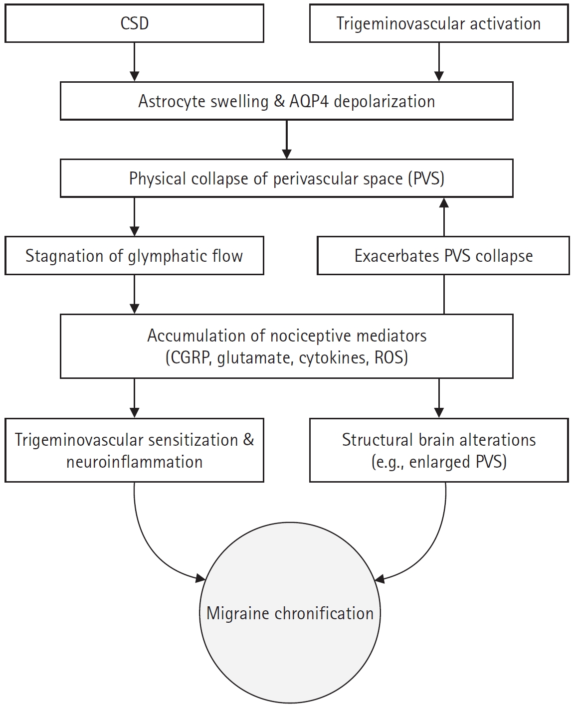


Figure 1.
Figure 2.
| Study (year) | Study design | MRI imaging modality | Study population (n) | Main findings |
|---|---|---|---|---|
| Dai et al.38 (2026) | Cross-sectional | DTI-ALPS & BOLD-CSF coupling | EM (n=200), CM (n=38), HC (n=65) | Significantly greater BAG and higher-order network abnormalities in CM compared to EM and HC. |
| Cho et al.40 (2026) | Retrospective case-control | MRI: ePVS visual rating | EM (n=44), CM (n=44), HC (n=39) | Higher prevalence of high-grade ePVS in CM compared to HC (in CSO and midbrain) and compared to EM (specifically in CSO). |
| Wang et al.24 (2025) | Cross-sectional | 7T DTI-ALPS & high-resolution structural imaging | MwA (n=35), MwoA (n=45), HC (n=39) | Prominent glymphatic dysfunction in MwA, evidenced by a lower ALPS index compared to HC and more enlarged PVS compared to MwoA. |
| Cai et al.33 (2025) | Cross-sectional | DTI-ALPS | EM (n=74), HC (n=42) | Significantly lower ALPS index in HFEM compared to LFEM and HC. |
| Ren et al.15 (2025) | Cross-sectional | DCE-MRI | EM (n=38), HC (n=22) | Impaired meningeal lymphatic drainage on DCE-MRI in EM compared to HC, which strongly correlates with headache intensity. |
| Wu et al.31 (2024) | Cross-sectional | DTI-ALPS | EM (n=73), CM (n=39), HC (n=63) | Significantly lower ALPS index in CM compared to EM and HC, with the most severe impairment observed in CM with MOH. |
| Zhang et al.36 (2023) | Cross-sectional | DTI-ALPS | EM (n=32) CM (n=24), HC (n=41) | The DTI-ALPS index was significantly higher in CM than EM and HC. |
| Cao et al.37 (2024) | Cross-sectional | DKI-ALPS | Migraine (n=37), HC (n=29) | Significantly higher DKI-ALPS index in migraine patients compared to HC. |
| Yuan et al.39 (2023) | Cross-sectional | ePVS visual rating | EM (n=59), CM (n=115), HC (n=57) | Significantly higher prevalence of high-grade ePVS in the CSO and midbrain in migraineurs compared to HC. |
| Ornello et al.35 (2023) | Cross-sectional | DTI-ALPS | Migraine (n=147) | White matter hyperintensity was not associated with an impairment in the glymphatic system in patients with migraine. |
| Lee et al.34 (2022) | Cross-sectional | DTI-ALPS | MwA (n=20), MwoA (n=72), HC (n=80) | The DTI-ALPS index did not differ between migraine patients and HC. |
| Study (year) | Experimental model | Target/mechanism assessed | Key findings |
|---|---|---|---|
| Pietra et al.30 (2026) | Mouse model of headache allodynia | α1-noradrenergic antagonism | Restoration of sleep-dependent glymphatic function and significant alleviation of migraine-like allodynia via α1-adrenergic receptor blockade. |
| Duan et al.20 (2025) | FHM2 mice | Neurovascular coupling | Severe glymphatic clearance impairment driven by abnormal neurovascular coupling and loss of AQP4 polarization. |
| Nelson-Maney et al.14 (2024) & Thomas et al.16 (2024) | Migraine mouse models (CGRP/NTG) | Meningeal lymphatic CGRP signaling | CGRP signaling decreases functional permeability of lymphatic vessels, hindering CSF drainage and driving neuroinflammation and pain. |
| Huang et al.12 (2023) | NTG-induced chronic migraine mice | AQP4 depolarization & Glymphatic flow | Severe glymphatic dysfunction and AQP4 depolarization, with exacerbated allodynia and CGRP accumulation upon AQP4 inhibition. |
| Mukherjee et al.19 (2023) | CFD model of CSD | Perivascular CSF flow dynamics | Prolonged suppression of perivascular CSF flow strongly coupled with the hemodynamic wave of spreading depolarization. |
| Rosic et al.21 (2019) | In vivo CSD model (Aqp4 KO mice) | Astroglial endfeet volume | Severe astrocyte endfoot swelling and PVS collapse during CSD, occurring robustly independent of AQP4 channels. |
| Enger et al.17 (2017) | Awake CSD mice (Aqp4 KO mice) | Extracellular Glutamate & Calcium signaling | Attenuated extracellular glutamate elevation during CSD, demonstrating a complex role of AQP4 in glutamate dynamics. |
| Schain et al.18 (2017) | In vivo two-photon imaging during CSD | Paravascular space closure | Rapid, sustained closure of the paravascular space during CSD, functionally arresting glymphatic CSF flow. |
MRI, magnetic resonance imaging; DTI, diffusion tensor imaging; ALPS, analysis along the perivascular space; BOLD, blood-oxygen-level-dependent; CSF, cerebrospinal fluid; EM, episodic migraine; CM, chronic migraine; HC, healthy controls; BAG, brain age gap; ePVS, enlarged perivascular spaces; CSO, centrum semiovale; MwA, migraine with aura; MwoA, migraine without aura; PVS, perivascular space; HFEM, high-frequency episodic migraine; LFEM, low-frequency episodic migraine; DCE, dynamic contrast-enhanced; MOH, medication-overuse headache; DKI, diffusion kurtosis imaging.
FHM2, familial hemiplegic migraine type 2; AQP4, aquaporin-4; CGRP, calcitonin gene-related peptide; NTG, nitroglycerin; CSF, cerebrospinal fluid; CFD, computational fluid dynamics; CSD, cortical spreading depression;
Table 1.
Table 2.
TOP


