
Articles
- Page Path
- HOME > Headache Pain Res > Ahead of print > Article
-
Review Article
Temporal Evolution and Multimodal Neuroimaging in Reversible Cerebral Vasoconstriction Syndrome, Arterial Dissection, and Cerebral Venous Thrombosis -
Young-Eun Gil

-
DOI: https://doi.org/10.62087/hpr.2026.0018
Published online: June 18, 2026
Department of Neurology, St. Vincent’s Hospital, College of Medicine, The Catholic University of Korea, Seoul, Republic of Korea
- Correspondence: Young-Eun Gil, M.D. Department of Neurology, St. Vincent’s Hospital, College of Medicine, The Catholic University of Korea, 93 Jungbu-daero, Paldal-gu, Suwon 16247, Republic of Korea Tel: +82-31-249-7170, Fax: +82-31-243-0306, E-mail: youngeungil.md@gmail.com
© 2026 The Korean Headache Society
This is an Open Access article distributed under the terms of the Creative Commons Attribution Non-Commercial License (https://creativecommons.org/licenses/by-nc/4.0) which permits unrestricted non-commercial use, distribution, and reproduction in any medium, provided the original work is properly cited.
- 46 Views
- 0 Download
Abstract
- Cerebrovascular disorders are important secondary causes of headache, but diagnosis can be challenging because headache may be the earliest or only presenting symptom and initial neuroimaging findings are often normal or nonspecific. This review provides a systematic, imaging-focused discussion of three representative cerebrovascular headache disorders: reversible cerebral vasoconstriction syndrome (RCVS), cervical and intracranial artery dissection, and cerebral venous thrombosis (CVT). For each condition, we describe characteristic findings across relevant imaging modalities, including computed tomography, computed tomography angiography, magnetic resonance imaging (MRI), magnetic resonance angiography, vessel wall (VW)-MRI, and susceptibility-sensitive sequences. We also discuss the temporal evolution of imaging findings, which underlies several common diagnostic pitfalls. In RCVS, angiographic vasoconstriction propagates centripetally from distal to proximal vessels, and contrast-enhanced fluid-attenuated inversion recovery imaging can detect blood–brain barrier disruption before vasoconstriction becomes angiographically apparent. In arterial dissection, VW-MRI can show mural features that confirm the diagnosis and may help stratify ischemic risk. In CVT, susceptibility-weighted imaging improves detection of cortical vein thrombosis, a subtype that can be missed on conventional venography. Across all three conditions, single-time-point imaging may be misleading, and serial imaging is often needed to increase diagnostic certainty because interval changes may reveal findings not present on the initial study. By integrating modality-specific findings with their temporal dynamics, this review proposes a practical imaging framework for the early and accurate diagnosis of cerebrovascular secondary headache disorders.
INTRODUCTION
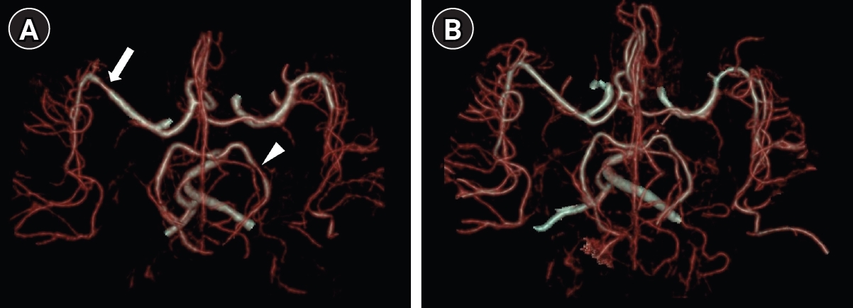
REVERSIBLE CEREBRAL VASOCONSTRICTION SYNDROME
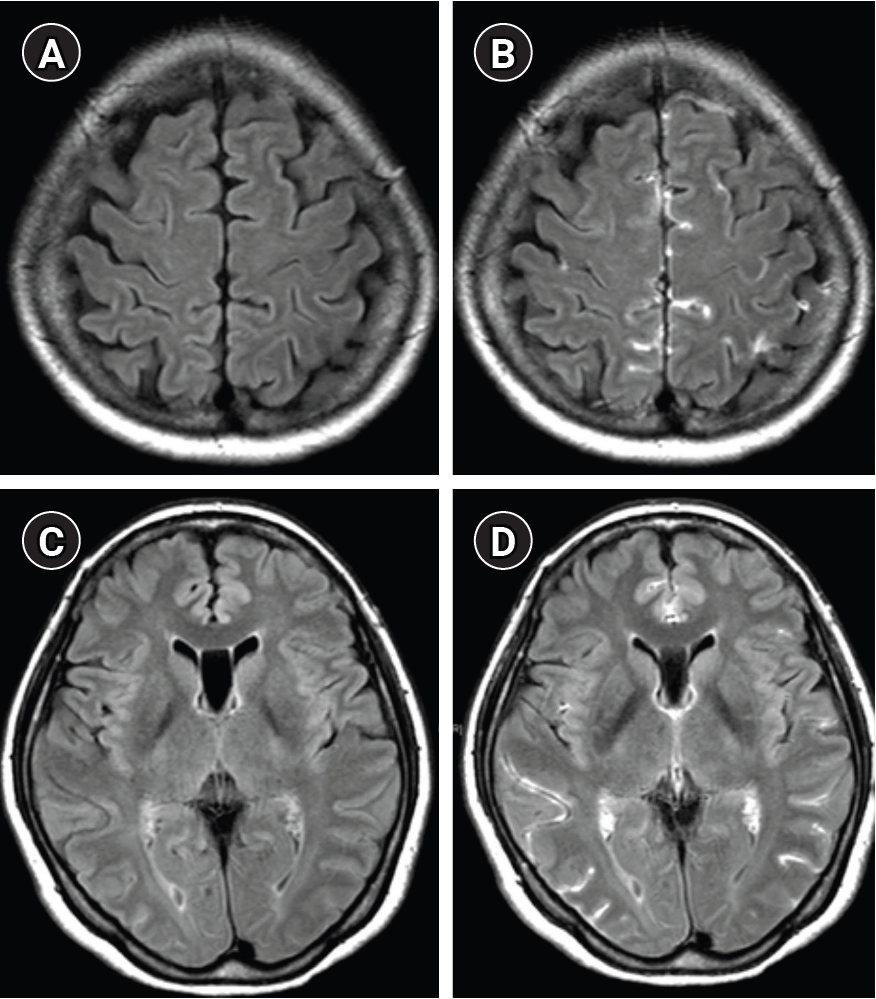
CERVICAL AND INTRACRANIAL ARTERY DISSECTION
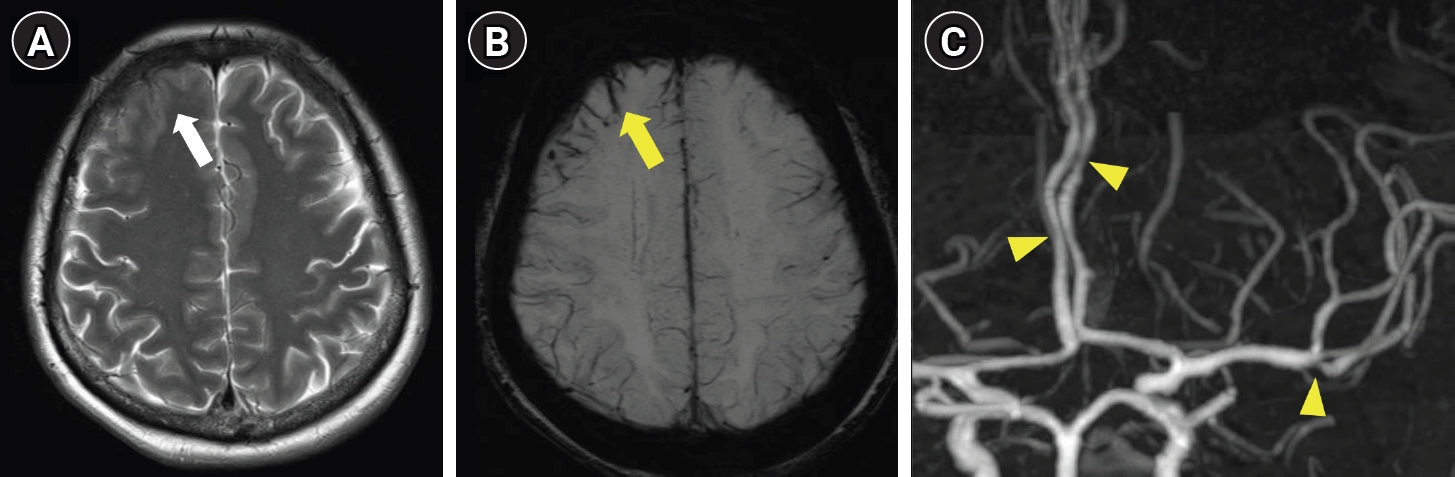
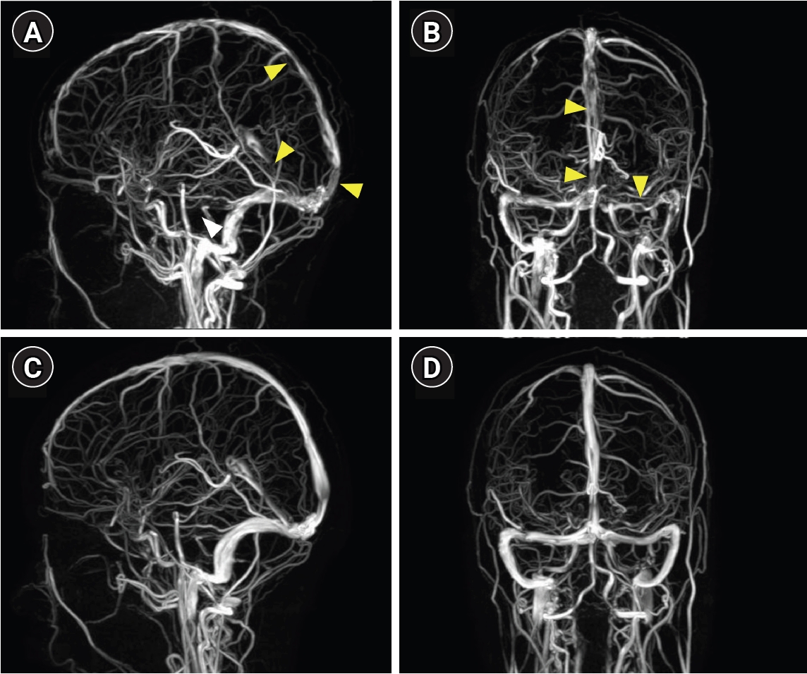
CEREBRAL VENOUS THROMBOSIS
INTEGRATED DIAGNOSTIC PEARLS AND PITFALLS
CONCLUSION
AVAILABILITY OF DATA AND MATERIAL
The data presented in this study are available upon reasonable request from the corresponding author.
AUTHOR CONTRIBUTIONS
Conceptualization: YEG; Writing–original draft: YEG; Writing–review & editing: YEG.
CONFLICT OF INTEREST
No potential conflict of interest relevant to this article was reported.
FUNDING STATEMENT
Not applicable.
ACKNOWLEDGMENTS
Not applicable.


| Feature | RCVS | PACNS | ICAD | Dissection |
|---|---|---|---|---|
| Wall thickening | Diffuse, circumferential | Diffuse, circumferential | Eccentric | Eccentric |
| Enhancement | None to mild* | Strong, persistent | Common (plaque) | Variable |
| T2 hyperintensity | Absent | Absent | Common | Variable (IMH) |
| Temporal behavior | Resolves<3 months | Persistent months–years | Stable | Evolves over weeks |
MRI, magnetic resonance imaging; RCVS, reversible cerebral vasoconstriction syndrome; PACNS, primary angiitis of the central nervous system; ICAD, intracranial atherosclerotic disease; IMH, intramural hematoma.
*Mild enhancement is present in a subset of RCVS patients and typically resolves on follow-up; this does not exclude the diagnosis. Strong or persistent enhancement should raise suspicion for PACNS rather than RCVS.13,14,33,34
RCVS, reversible cerebral vasoconstriction syndrome; CVT, cerebral venous thrombosis; CTA, computed tomography angiography; MRA, magnetic resonance angiography; CE-FLAIR, contrast-enhanced fluid-attenuated inversion recovery; BBB, blood–brain barrier; MRI, magnetic resonance imaging; CT, computed tomography; CTV, computed tomography venography; MRV, magnetic resonance venography; SWI, susceptibility-weighted imaging; GRE, gradient-echo.
RCVS, reversible cerebral vasoconstriction syndrome; CVT, cerebral venous thrombosis; CE-FLAIR, contrast-enhanced fluid-attenuated inversion recovery; BBB, blood–brain barrier; SAH, subarachnoid hemorrhage; CTA, computed tomography angiography; MRA, magnetic resonance angiography; ICAD, intracranial atherosclerotic disease; MRI, magnetic resonance imaging; VW-MRI, vessel wall magnetic resonance imaging; DWI, diffusion-weighted imaging; CT, computed tomography; MRV, magnetic resonance venography; CTV, computed tomography venography; SWI, susceptibility-weighted imaging; GRE, gradient-echo; MR, magnetic resonance.
- 1. GBD 2023 Headache Collaborators. Global, regional, and national burden of headache disorders, 1990-2023: a systematic analysis for the Global Burden of Disease Study 2023. Lancet Neurol 2025;24:1005-1015.ArticlePubMedPMC
- 2. GBD 2023 Disease and Injury and Risk Factor Collaborators. Burden of 375 diseases and injuries, risk-attributable burden of 88 risk factors, and healthy life expectancy in 204 countries and territories, including 660 subnational locations, 1990-2023: a systematic analysis for the Global Burden of Disease Study 2023. Lancet 2025;406:1873-1922.ArticlePubMedPMC
- 3. Headache Classification Committee of the International Headache Society (IHS). The International Classification of Headache Disorders, 3rd edition. Cephalalgia 2018;38:1-211.ArticlePDF
- 4. Do TP, Remmers A, Schytz HW, et al. Red and orange flags for secondary headaches in clinical practice: SNNOOP10 list. Neurology 2019;92:134-144.ArticlePubMed
- 5. Kim JS. Headache and stroke: a review. Headache Pain Res 2026;27:7-12.ArticlePDF
- 6. Ducros A, Wolff V. The typical thunderclap headache of reversible cerebral vasoconstriction syndrome and its various triggers. Headache 2016;56:657-673.ArticlePubMed
- 7. Ducros A. Reversible cerebral vasoconstriction syndrome. Lancet Neurol 2012;11:906-917.ArticlePubMed
- 8. Doukhi D, Debette S, Mawet J. Headaches attributed to cranial and cervical artery dissections. J Headache Pain 2025;26:28.ArticlePubMedPMCPDF
- 9. Cumurciuc R, Crassard I, Sarov M, Valade D, Bousser MG. Headache as the only neurological sign of cerebral venous thrombosis: a series of 17 cases. J Neurol Neurosurg Psychiatry 2005;76:1084-1087.ArticlePubMedPMC
- 10. Rodallec MH, Marteau V, Gerber S, Desmottes L, Zins M. Craniocervical arterial dissection: spectrum of imaging findings and differential diagnosis. Radiographics 2008;28:1711-1728.ArticlePubMed
- 11. Leach JL, Fortuna RB, Jones BV, Gaskill-Shipley MF. Imaging of cerebral venous thrombosis: current techniques, spectrum of findings, and diagnostic pitfalls. Radiographics 2006;26 Suppl 1:S19-S43.ArticlePubMed
- 12. Calabrese LH, Dodick DW, Schwedt TJ, Singhal AB. Narrative review: reversible cerebral vasoconstriction syndromes. Ann Intern Med 2007;146:34-44.ArticlePubMedPDF
- 13. Obusez EC, Hui F, Hajj-Ali RA, et al. High-resolution MRI vessel wall imaging: spatial and temporal patterns of reversible cerebral vasoconstriction syndrome and central nervous system vasculitis. AJNR Am J Neuroradiol 2014;35:1527-1532.ArticlePubMedPMC
- 14. Chen CY, Chen SP, Fuh JL, et al. Vascular wall imaging in reversible cerebral vasoconstriction syndrome: a 3-T contrast-enhanced MRI study. J Headache Pain 2018;19:74.ArticlePubMedPMCPDF
- 15. Chen SP, Fuh JL, Lirng JF, Wang YF, Wang SJ. Recurrence of reversible cerebral vasoconstriction syndrome: a long-term follow-up study. Neurology 2015;84:1552-1558.ArticlePubMed
- 16. Boitet R, de Gaalon S, Duflos C, et al. Long-term outcomes after reversible cerebral vasoconstriction syndrome. Stroke 2020;51:670-673.ArticlePubMed
- 17. Ducros A, Boukobza M, Porcher R, Sarov M, Valade D, Bousser MG. The clinical and radiological spectrum of reversible cerebral vasoconstriction syndrome. A prospective series of 67 patients. Brain 2007;130:3091-3101.ArticlePubMed
- 18. Singhal AB, Hajj-Ali RA, Topcuoglu MA, et al. Reversible cerebral vasoconstriction syndromes: analysis of 139 cases. Arch Neurol 2011;68:1005-1012.ArticlePubMed
- 19. Choi HA, Lee MJ, Choi H, Chung CS. Characteristics and demographics of reversible cerebral vasoconstriction syndrome: a large prospective series of Korean patients. Cephalalgia 2018;38:765-775.ArticlePubMedPDF
- 20. Magid-Bernstein J, Omran SS, Parikh NS, Merkler AE, Navi B, Kamel H. Reversible cerebral vasoconstriction syndrome: symptoms, incidence, and resource utilization in a population-based US cohort. Neurology 2021;97:e248-e253.ArticlePubMedPMC
- 21. Wolff V, Ducros A. Reversible cerebral vasoconstriction syndrome without typical thunderclap headache. Headache 2016;56:674-687.ArticlePubMed
- 22. Lange KS, Wang SJ, Pezzini A, et al. Regional differences in presentation, cause, and outcome of reversible cerebral vasoconstriction syndrome. Stroke 2025;56:2516-2526.ArticlePubMed
- 23. Singhal AB, Topcuoglu MA, Fok JW, et al. Reversible cerebral vasoconstriction syndromes and primary angiitis of the central nervous system: clinical, imaging, and angiographic comparison. Ann Neurol 2016;79:882-894.ArticlePubMedPDF
- 24. Miller TR, Shivashankar R, Mossa-Basha M, Gandhi D. Reversible cerebral vasoconstriction syndrome, part 2: diagnostic work-up, imaging evaluation, and differential diagnosis. AJNR Am J Neuroradiol 2015;36:1580-1588.ArticlePubMedPMC
- 25. Ducros A, Bousser MG. Reversible cerebral vasoconstriction syndrome. Pract Neurol 2009;9:256-267.ArticlePubMed
- 26. Chen SP, Fuh JL, Wang SJ, et al. Magnetic resonance angiography in reversible cerebral vasoconstriction syndromes. Ann Neurol 2010;67:648-656.ArticlePubMed
- 27. Edelman RR, Koktzoglou I. Noncontrast MR angiography: an update. J Magn Reson Imaging 2019;49:355-373.ArticlePubMedPDF
- 28. Katz BS, Fugate JE, Ameriso SF, et al. Clinical worsening in reversible cerebral vasoconstriction syndrome. JAMA Neurol 2014;71:68-73.ArticlePubMed
- 29. Kim SA, Kim EY, Wang SJ, Lee MJ. Beyond the “string of beads”: case-based exploration of diagnostic pitfalls and solutions in reversible cerebral vasoconstriction syndrome. J Headache Pain 2025;26:89.ArticlePubMedPMCPDF
- 30. Topcuoglu MA, Singhal AB. Hemorrhagic reversible cerebral vasoconstriction syndrome: features and mechanisms. Stroke 2016;47:1742-1747.ArticlePubMed
- 31. Ducros A, Fiedler U, Porcher R, Boukobza M, Stapf C, Bousser MG. Hemorrhagic manifestations of reversible cerebral vasoconstriction syndrome: frequency, features, and risk factors. Stroke 2010;41:2505-2511.ArticlePubMed
- 32. Pilato F, Distefano M, Calandrelli R. Posterior reversible encephalopathy syndrome and reversible cerebral vasoconstriction syndrome: clinical and radiological considerations. Front Neurol 2020;11:34.ArticlePubMedPMC
- 33. Mandell DM, Mossa-Basha M, Qiao Y, et al. Intracranial vessel wall MRI: principles and expert consensus recommendations of the American Society of Neuroradiology. AJNR Am J Neuroradiol 2017;38:218-229.ArticlePubMed
- 34. Mossa-Basha M, Hwang WD, De Havenon A, et al. Multicontrast high-resolution vessel wall magnetic resonance imaging and its value in differentiating intracranial vasculopathic processes. Stroke 2015;46:1567-1573.ArticlePubMed
- 35. Shimoda M, Oda S, Hirayama A, et al. Centripetal propagation of vasoconstriction at the time of headache resolution in patients with reversible cerebral vasoconstriction syndrome. AJNR Am J Neuroradiol 2016;37:1594-1598.ArticlePubMedPMC
- 36. Cho S, Ling YH, Lee MJ, et al. Temporal profile of blood-brain barrier breakdown in reversible cerebral vasoconstriction syndrome. Stroke 2020;51:1451-1457.ArticlePubMed
- 37. Lee MJ, Cha J, Choi HA, et al. Blood-brain barrier breakdown in reversible cerebral vasoconstriction syndrome: implications for pathophysiology and diagnosis. Ann Neurol 2017;81:454-466.ArticlePubMedPDF
- 38. Rocha EA, Topcuoglu MA, Silva GS, Singhal AB. RCVS2 score and diagnostic approach for reversible cerebral vasoconstriction syndrome. Neurology 2019;92:e639-e647.ArticlePubMed
- 39. Cho S, Lee MJ, Gil YE, Chung CS. RCVS-TCH score can predict reversible cerebral vasoconstriction syndrome in patients with thunderclap headache. Sci Rep 2021;11:7750.ArticlePubMedPMCPDF
- 40. Godwin SA, Cherkas DS, Panagos PD, Shih RD, Byyny R, Wolf SJ; American College of Emergency Physicians Clinical Policies Subcommittee (Writing Committee) on Acute Headache. Clinical policy: critical issues in the evaluation and management of adult patients presenting to the emergency department with acute headache. Ann Emerg Med 2019;74:e41-e74.ArticlePubMed
- 41. Long D, Koyfman A, Long B. The thunderclap headache: approach and management in the emergency department. J Emerg Med 2019;56:633-641.ArticlePubMed
- 42. Perry JJ, Stiell IG, Sivilotti ML, et al. Sensitivity of computed tomography performed within six hours of onset of headache for diagnosis of subarachnoid haemorrhage: prospective cohort study. BMJ 2011;343:d4277.ArticlePubMedPMC
- 43. Debette S, Leys D. Cervical-artery dissections: predisposing factors, diagnosis, and outcome. Lancet Neurol 2009;8:668-678.ArticlePubMed
- 44. Griffin KJ, Harmsen WS, Mandrekar J, Brown RD Jr, Keser Z. Epidemiology of spontaneous cervical artery dissection: population-based study. Stroke 2024;55:670-677.ArticlePubMed
- 45. Keser Z, Chiang CC, Benson JC, Pezzini A, Lanzino G. Cervical artery dissections: etiopathogenesis and management. Vasc Health Risk Manag 2022;18:685-700.ArticlePubMedPMCPDF
- 46. Debette S, Compter A, Labeyrie MA, et al. Epidemiology, pathophysiology, diagnosis, and management of intracranial artery dissection. Lancet Neurol 2015;14:640-654.ArticlePubMed
- 47. Arnold M, Cumurciuc R, Stapf C, Favrole P, Berthet K, Bousser MG. Pain as the only symptom of cervical artery dissection. J Neurol Neurosurg Psychiatry 2006;77:1021-1024.ArticlePubMedPMC
- 48. Vertinsky AT, Schwartz NE, Fischbein NJ, Rosenberg J, Albers GW, Zaharchuk G. Comparison of multidetector CT angiography and MR imaging of cervical artery dissection. AJNR Am J Neuroradiol 2008;29:1753-1760.ArticlePubMedPMC
- 49. Wu Y, Wu F, Liu Y, et al. High-resolution magnetic resonance imaging of cervicocranial artery dissection: imaging features associated with stroke. Stroke 2019;50:3101-3107.ArticlePubMedPMC
- 50. Morel A, Naggara O, Touzé E, et al. Mechanism of ischemic infarct in spontaneous cervical artery dissection. Stroke 2012;43:1354-1361.ArticlePubMed
- 51. Debette S, Mazighi M, Bijlenga P, et al. ESO guideline for the management of extracranial and intracranial artery dissection. Eur Stroke J 2021;6:XXXIX-LXXXVIII.ArticlePubMedPMCPDF
- 52. Yaghi S, Engelter S, Del Brutto VJ, et al. Treatment and outcomes of cervical artery dissection in adults: a scientific statement from the American Heart Association. Stroke 2024;55:e91-e106.ArticlePubMed
- 53. Saposnik G, Bushnell C, Coutinho JM, et al. Diagnosis and management of cerebral venous thrombosis: a scientific statement from the American Heart Association. Stroke 2024;55:e77-e90.ArticlePubMed
- 54. Canedo-Antelo M, Baleato-González S, Mosqueira AJ, et al. Radiologic clues to cerebral venous thrombosis. Radiographics 2019;39:1611-1628.ArticlePubMed
- 55. Ferro JM, Canhão P, Stam J, Bousser MG, Barinagarrementeria F; ISCVT Investigators. Prognosis of cerebral vein and dural sinus thrombosis: results of the International Study on Cerebral Vein and Dural Sinus Thrombosis (ISCVT). Stroke 2004;35:664-670.ArticlePubMed
- 56. Idbaih A, Boukobza M, Crassard I, Porcher R, Bousser MG, Chabriat H. MRI of clot in cerebral venous thrombosis: high diagnostic value of susceptibility-weighted images. Stroke 2006;37:991-995.ArticlePubMed
- 57. Coutinho JM, Gerritsma JJ, Zuurbier SM, Stam J. Isolated cortical vein thrombosis: systematic review of case reports and case series. Stroke 2014;45:1836-1838.ArticlePubMed
- 58. Boukobza M, Crassard I, Bousser MG, Chabriat H. MR imaging features of isolated cortical vein thrombosis: diagnosis and follow-up. AJNR Am J Neuroradiol 2009;30:344-348.ArticlePubMed
- 59. Mullins ME, Grant PE, Wang B, Gonzalez RG, Schaefer PW. Parenchymal abnormalities associated with cerebral venous sinus thrombosis: assessment with diffusion-weighted MR imaging. AJNR Am J Neuroradiol 2004;25:1666-1675.PubMedPMCPDF
- 60. Stolz E, Trittmacher S, Rahimi A, et al. Influence of recanalization on outcome in dural sinus thrombosis: a prospective study. Stroke 2004;35:544-547.ArticlePubMed
REFERENCES
Figure & Data
References
Citations
 PubReader
 ePub Link
-
 Cite this Article
- Cite this Article
-
- Close
- Download Citation
- Close
- Figure
-
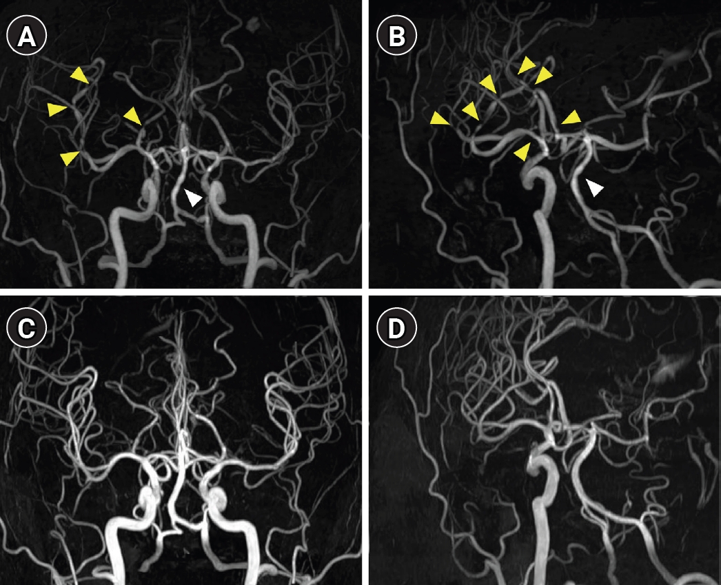
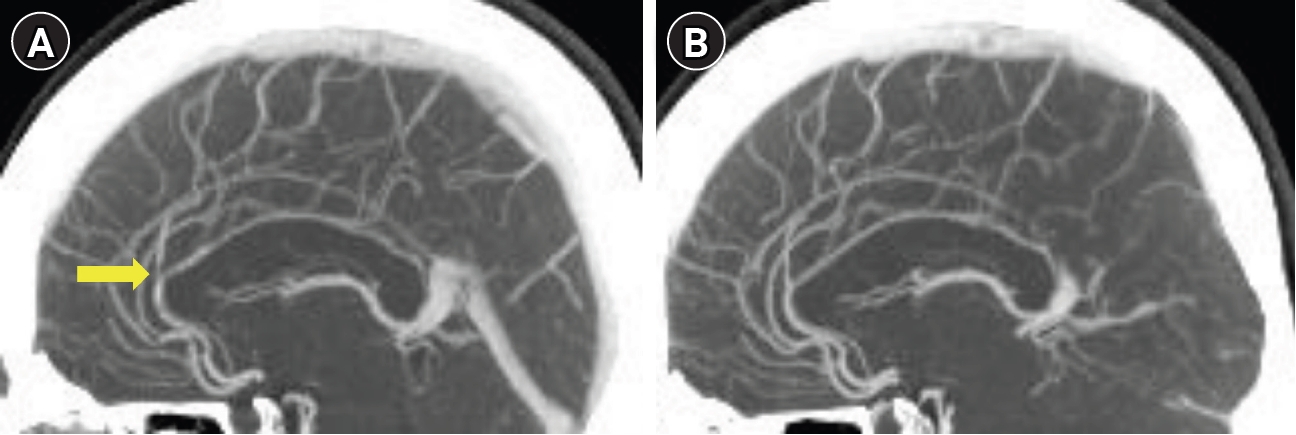

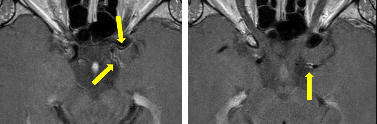
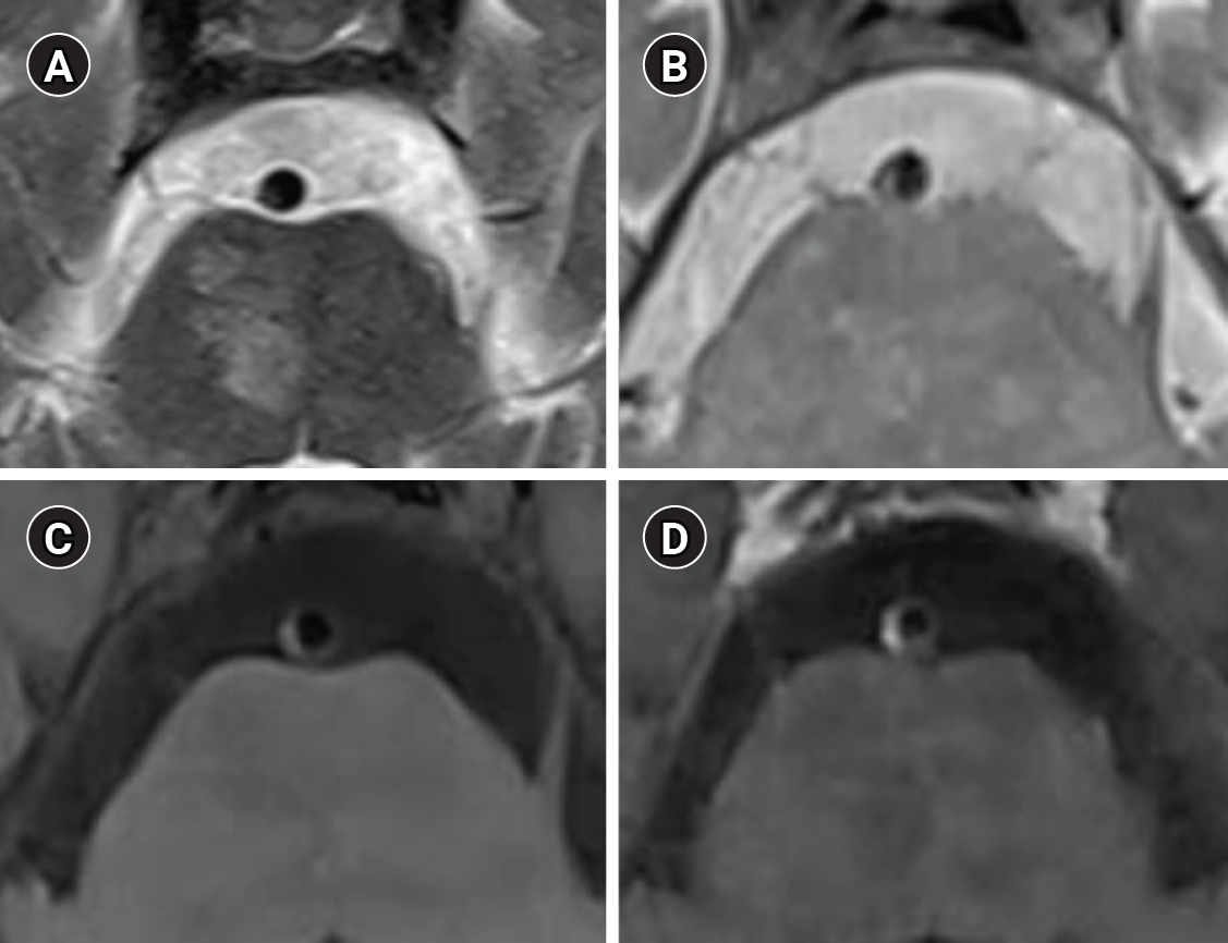
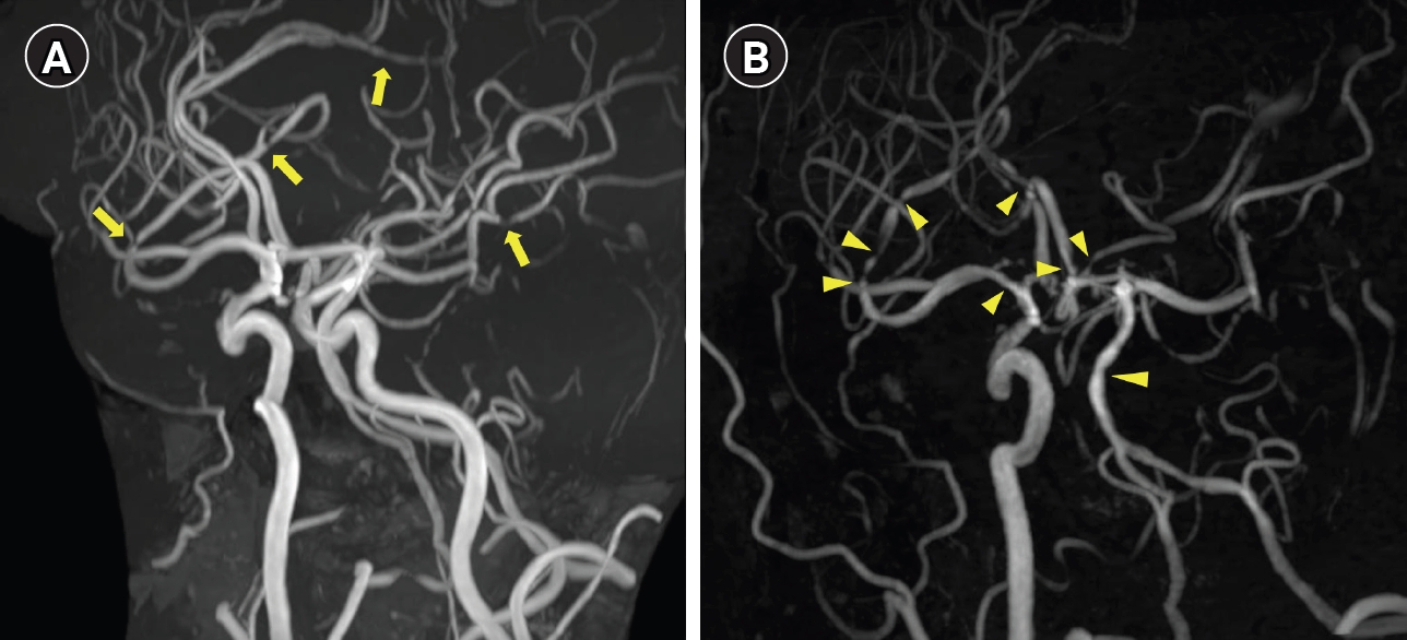


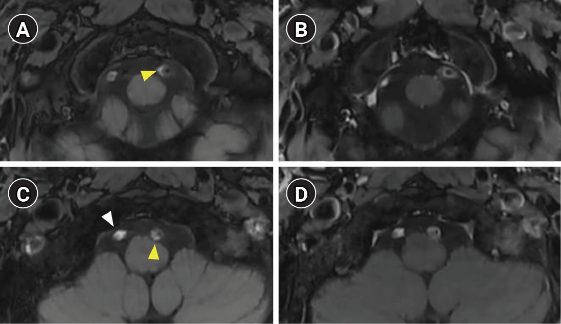
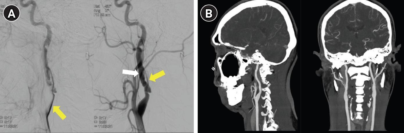
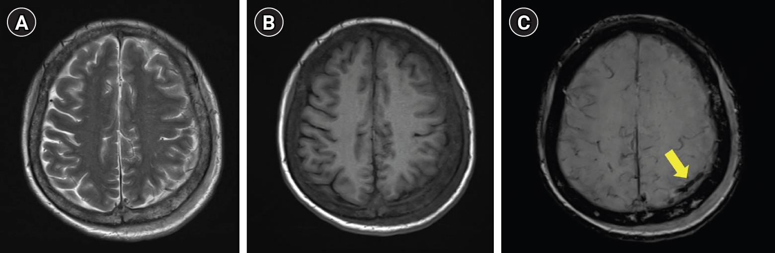


Figure 1.
Figure 2.
Figure 3.
Figure 4.
Figure 5.
Figure 6.
Figure 7.
Figure 8.
Figure 9.
Figure 10.
Figure 11.
Figure 12.
| Feature | RCVS | PACNS | ICAD | Dissection |
|---|---|---|---|---|
| Wall thickening | Diffuse, circumferential | Diffuse, circumferential | Eccentric | Eccentric |
| Enhancement | None to mild |
Strong, persistent | Common (plaque) | Variable |
| T2 hyperintensity | Absent | Absent | Common | Variable (IMH) |
| Temporal behavior | Resolves<3 months | Persistent months–years | Stable | Evolves over weeks |
| Disease | Initial imaging pitfall | Subacute evolution (days to weeks) | Follow-up evolution | Practical implication |
|---|---|---|---|---|
| RCVS | CTA/MRA may be normal (~21%, 1st week); CE-FLAIR may show BBB breakdown (±vasoconstriction) | Centripetal propagation (distal → proximal); “string of beads“ most prominent at 1–3 weeks (peak, ~day 16) | Reversibility by ~3 months in most patients; delayed or incomplete resolution in a subset | Repeat vascular imaging at 1–2 weeks if initial unremarkable; reversibility on follow-up required for definite diagnosis |
| Arterial dissection | Intramural hematoma may be T1-isointense in early phase (false-negative MRI); luminal study—subtle tapering only | T1 hyperintensity emerges (methemoglobin); eccentric hematoma, intimal flap, luminal irregularity become more conspicuous | Healing/remodeling in most cases (3–6 months); residual stenosis or pseudoaneurysm may persist | Repeat imaging within days–weeks if early study equivocal; tailor follow-up modality to clinical question (MRI/MRA for hematoma resolution; CTA for residual stenosis/pseudoaneurysm) |
| CVT | Noncontrast CT may be normal; conventional MRI thrombus signal may mimic flow void; cortical vein thrombosis is often missed on CTV/MRV alone | SWI/GRE—prominent blooming (deoxyhemoglobin); venous infarction±hemorrhage may develop | Recanalization over weeks–months on anticoagulation; partial patency in subset | SWI or GRE should be routinely included for suspected CVT; follow-up MRV after anticoagulation to document recanalization |
| Disease | Clinical context | Key imaging pearl | Major pitfall | Value of follow-up imaging |
|---|---|---|---|---|
| RCVS | Recurrent triggered thunderclap headache; initial vascular imaging may be normal. | Multifocal vasoconstriction with distal-to-proximal (centripetal) evolution; CE-FLAIR may show BBB breakdown before angiographic peak; bilateral cortical-subcortical border-zone infarcts or convexal SAH can support the diagnosis. | Early CTA/MRA may be false-negative (~21%); long-segment or non-beaded stenosis may mimic ICAD or vasculitis; clinical improvement does not parallel angiographic recovery. | Repeat CTA/MRA at 1–2 weeks may reveal interval worsening; later reversibility supports the diagnosis, although delayed or incomplete resolution occurs in a subset. |
| Arterial dissection | New unilateral headache or neck pain, sometimes preceding stroke; thunderclap onset can occur. | CTA shows tapering stenosis, intimal flap, or pseudoaneurysm; MRI/VW-MRI demonstrates eccentric intramural hematoma or intimal injury; DWI detects associated ischemia. | Hyperacute (<48 hr) mural hematoma may be T1 iso-/hypointense; subtle luminal change may mimic atherosclerotic stenosis; pain may be the only manifestation. | Serial CTA/MRA or VW-MRI helps confirm remodeling, persistent stenosis, or pseudoaneurysm and clarifies initially equivocal lesions. |
| CVT | Headache-predominant presentation; noncontrast CT may be normal or nonspecific. | MRI with MRV/CTV improves diagnosis; SWI/GRE is particularly useful for cortical vein thrombosis; venous edema or hemorrhage may cross arterial territories. | Noncontrast CT may be normal; conventional MR signal may mimic a flow void; MRV/CTV alone may miss isolated cortical vein thrombosis. | Follow-up imaging documents recanalization and helps distinguish reversible venous edema from irreversible venous infarction. |
MRI, magnetic resonance imaging; RCVS, reversible cerebral vasoconstriction syndrome; PACNS, primary angiitis of the central nervous system; ICAD, intracranial atherosclerotic disease; IMH, intramural hematoma. Mild enhancement is present in a subset of RCVS patients and typically resolves on follow-up; this does not exclude the diagnosis. Strong or persistent enhancement should raise suspicion for PACNS rather than RCVS.
RCVS, reversible cerebral vasoconstriction syndrome; CVT, cerebral venous thrombosis; CTA, computed tomography angiography; MRA, magnetic resonance angiography; CE-FLAIR, contrast-enhanced fluid-attenuated inversion recovery; BBB, blood–brain barrier; MRI, magnetic resonance imaging; CT, computed tomography; CTV, computed tomography venography; MRV, magnetic resonance venography; SWI, susceptibility-weighted imaging; GRE, gradient-echo.
RCVS, reversible cerebral vasoconstriction syndrome; CVT, cerebral venous thrombosis; CE-FLAIR, contrast-enhanced fluid-attenuated inversion recovery; BBB, blood–brain barrier; SAH, subarachnoid hemorrhage; CTA, computed tomography angiography; MRA, magnetic resonance angiography; ICAD, intracranial atherosclerotic disease; MRI, magnetic resonance imaging; VW-MRI, vessel wall magnetic resonance imaging; DWI, diffusion-weighted imaging; CT, computed tomography; MRV, magnetic resonance venography; CTV, computed tomography venography; SWI, susceptibility-weighted imaging; GRE, gradient-echo; MR, magnetic resonance.
Table 1.
Table 2.
Table 3.
TOP

