Spontaneous Intracranial Hypotension Masquerading as Headache from an Unruptured Middle Cerebral Artery Aneurysm
Article information

Abstract
Spontaneous intracranial hypotension (SIH) is characterized by neurological symptoms resulting from cerebrospinal fluid leakage or volume depletion. Although orthostatic headache is the hallmark symptom, SIH can also present with nausea, vomiting, dizziness, tinnitus, and other neurological symptoms. In this case, a cerebral aneurysm identified during evaluation of the headache was initially considered a potential cause, prompting a decision to treat it. However, an orthostatic headache severe enough to interfere with standing or physical activity had been overlooked. After a pre-existing subdural effusion was identified, the evolving orthostatic features of the headache and the imaging findings raised suspicion of SIH. The headache resolved after a lumbar epidural blood patch. Although a cerebral aneurysm is a potential cause of headache, SIH may be difficult to recognize early. Careful history taking, particularly about the characteristics and postural nature of the headache, is therefore essential to appropriately evaluate patients with headache.
INTRODUCTION
Spontaneous intracranial hypotension (SIH) is a condition characterized by orthostatic headache that worsens when standing and improves when lying down, typically caused by spontaneous spinal cerebrospinal fluid (CSF) leakage or CSF hypotension. In addition, various other symptoms may occur, including nausea, vomiting, neck pain, neck stiffness, other ear-related symptoms, hearing disturbances, dizziness, and tinnitus.1 According to the International Classification of Headache Disorders, third edition, SIH is diagnosed when it is associated with a spontaneous, imaging-verified CSF leak and/or a lumbar puncture opening pressure <60 mm CSF.2
SIH is a disease with high rates of misdiagnosis and underdiagnosis.3 While orthostatic headache is a characteristic symptom, many patients also complain of other symptoms. Furthermore, its clear pathophysiological mechanism has not been elucidated, and it is a disease that is unfamiliar in clinical practice.4 Due to these various factors, the diagnosis of SIH is often delayed or misdiagnosed as other conditions.
In this case report, we describe a patient in whom a cerebral aneurysm was identified during the evaluation of headache and was initially considered a potential cause, while the possibility of SIH was not recognized early. This case highlights the importance of careful history taking, particularly regarding the characteristics and postural nature of headache, when evaluating its underlying causes.
CASE REPORT
A 47-year-old male presented with a gradually developing headache involving the entire head, which progressively worsened over time. There was no history of known connective tissue disorders or recent minor trauma preceding the onset of the headache. A clear orthostatic pattern was not documented at the initial presentation. The patient did not report nausea, vomiting, neck pain or stiffness, auditory symptoms, dizziness, or tinnitus. Brain high-resolution magnetic resonance imaging (MRI) and magnetic resonance angiography performed approximately 1 week after the onset of the headache revealed a 7-mm aneurysm at the bifurcation of the left middle cerebral artery (MCA). Retrospective review of the initial brain MRI from an SIH-focused perspective revealed no definite brain sagging or venous distension. Pachymeningeal enhancement could not be assessed because contrast-enhanced images were not obtained. The aneurysm was considered for preventive treatment independently of the presenting headache, based on its size, MCA bifurcation location, and the patient’s relatively young age after neurosurgical evaluation. During the 1-month waiting period for surgery, the patient took a leave of absence from work and spent most of the time in a supine position due to severe headaches that were exacerbated by standing or movement. A brain computed tomography (CT) angiography performed the day before the scheduled surgery confirmed the presence of the aneurysm (Figure 1). However, during the surgery, an unexpected subdural effusion was discovered. A consultation with the neurology department was requested immediately, raising suspicion of SIH as an important contributor to the patient's headache. A brain CT scan performed immediately after the clipping procedure revealed a subdural effusion. A careful retrospective review of the CT angiography obtained the day before surgery revealed that the subdural effusion had already been present at that time but had not been recognized initially (Figure 2A, B). Because SIH had not been recognized before the perioperative identification of the subdural effusion, specific conservative treatment for SIH had not been attempted. Approximately 10 days after clipping, the patient underwent a single non-targeted lumbar epidural blood patch (EBP) at the L2–L3 intervertebral level under imaging guidance, with injection of 20 mL of autologous blood. Following the EBP, the headache resolved. Follow-up brain CT performed 2 months later showed complete resolution of the subdural effusion (Figure 2C).
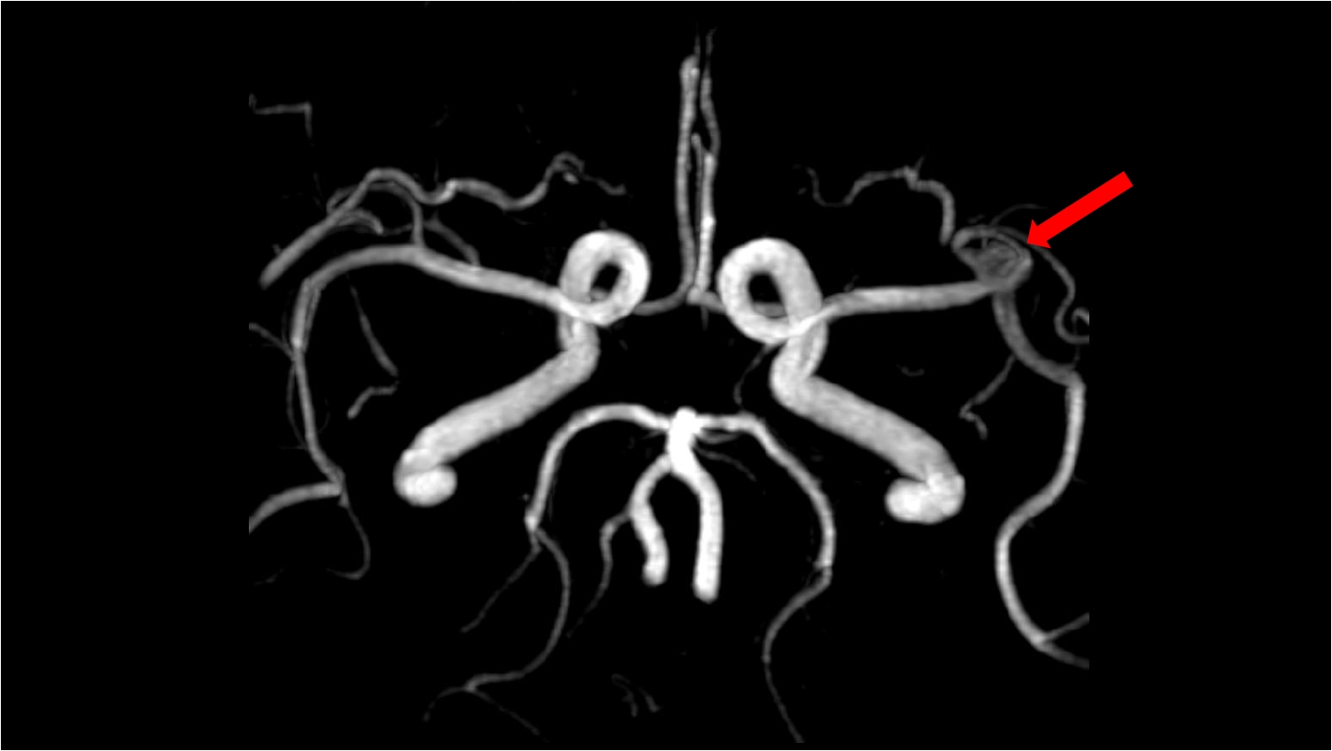
Cerebral aneurysm at the left middle cerebral artery bifurcation on brain computed tomography (CT) angiography. Brain CT angiography shows a 7-mm aneurysm (arrow) at the left middle cerebral artery bifurcation.
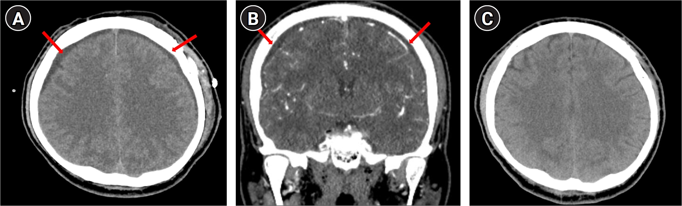
Serial brain computed tomography (CT) images showing changes in the subdural effusion. (A) Brain CT obtained immediately after aneurysm clipping shows bilateral subdural effusions (arrows). (B) Retrospective review of a coronal multiplanar reconstruction from CT angiography performed 1 day before surgery shows that the subdural effusion (arrows) was already present preoperatively. (C) Follow-up brain CT obtained 2 months after the epidural blood patch shows complete resolution of the subdural effusion.
DISCUSSION
This case illustrates a delayed diagnosis of SIH, in which a co-existing cerebral aneurysm was initially considered the cause of the headache despite the presence of a severe orthostatic headache. SIH was suspected after a subdural effusion was identified during surgery. Retrospective review of the CT angiography obtained before surgery revealed that the subdural effusion had already been present but had not been recognized at the time. Although the clinical presentation of SIH in this patient eventually became relatively typical, the characteristic clinical clue was not sufficiently incorporated into the diagnostic reasoning after an unruptured intracranial aneurysm (UIA) was identified. This case highlights the importance of considering alternative causes of headache even when a potentially relevant structural lesion is identified.
SIH is a clinical and radiological condition caused by spinal CSF leakage. This is due to a dural tear, leaking meningeal diverticulum or CSF venous fistula. The estimated incidence of SIH in the USA is 3.8 per 100,000 population per year.5,6 The most common symptom of SIH is orthostatic headache, which occurs or worsens when standing and improves when lying down. This symptom is observed in approximately 92% of SIH patients.1 Cases without orthostatic headache are observed in 5%, and cases without headache at all are observed in 3%. In our case, a clear orthostatic pattern was not documented at the initial presentation but became apparent as the headache progressively worsened. Despite the subsequent development of this characteristic clinical feature, the possibility of SIH was not sufficiently considered. In actual clinical practice, diagnosis of SIH is often delayed or misdiagnosed due to a lack of awareness of the disease and limited access to tests such as myelography or blood patch therapy.7,8
UIAs are commonly observed in the general population, found in approximately 3.2% of the global adult population (average age 50).9 Recently, due to the widespread use of high-resolution MRI, the frequency of incidental discovery is increasing.9 When an aneurysm is found in a patient with a headache, consequently, alternative causes of the headache were not sufficiently investigated. Although headache is frequently reported in patients with UIA, whether the aneurysm itself is responsible for the headache remains uncertain.10 However, the majority of UIAs will not rupture. For example, about 32,000 people out of a million in the general population with an average age of 50 have UIA, but it is estimated that only 0.25% of them (1 in 400 people) will rupture. In this case, the co-existing UIA was initially considered the cause of the headache despite the uncertain causal relationship between UIA and headache. Consequently, alternative causes of the headache were not sufficiently investigated, leading to a delay in the diagnosis of SIH.
SIH can be accompanied by various severe complications. These include subdural hematoma, subdural effusion, cerebral venous sinus thrombosis, superficial siderosis, bibrachial amyotrophy, syringomyelia secondary to pseudo-Chiari malformation, brain sagging dementia, and coma.8,11-13 SIH-related brain sagging and compensatory intracranial volume changes may result in subdural fluid collections, while traction on bridging veins may particularly contribute to subdural hematoma formation. The subdural effusion observed in this case was considered to have occurred through the same mechanism. Although craniotomy itself may contribute to postoperative subdural fluid collection, the subdural effusion in this patient was already present on CT angiography obtained 1 day before surgery. Moreover, comparison of the preoperative and immediate postoperative images showed no apparent increase in the extent of the effusion after surgery. Thus, although a contribution of the surgical procedure cannot be completely excluded, there was no radiological evidence of substantial aggravation of the pre-existing subdural effusion following surgery.
Blood patches have been applied to SIH since a relatively early period and remain a central treatment for SIH to this day.1,14 The mechanism of the blood patch involves the temporary increase in volume and pressure caused by the injection of blood into the epidural space, and the effect of the injected blood moving to the dural defect site to directly seal the leak.15,16 In our case as well, the headache improved after blood patch treatment, allowing the patient to be discharged.
A limitation of this case is the absence of confirmatory spinal imaging. Therefore, the site and mechanism of a spinal CSF leak could not be established. Nevertheless, the diagnosis of SIH was strongly supported by the progressive orthostatic headache, preoperative bilateral subdural effusion, and sustained clinical and radiological improvement following lumbar EBP. This case demonstrates the importance of not attributing headache solely to a co-existing cerebral aneurysm and of carefully considering clinical clues, particularly the orthostatic nature of the headache, and relevant imaging findings to avoid overlooking alternative causes such as SIH.
Notes
AVAILABILITY OF DATA AND MATERIAL
The data presented in this study are available upon reasonable request from the corresponding author.
AUTHOR CONTRIBUTIONS
Conceptualization: MJC, JK; Data curation: MJC, JK; Investigation: MJC, JK; Methodology: MJC, JK; Software: MJC, JK; Supervision: MJC, JK; Validation: MJC, JK; Visualization: MJC, JK; Writing–original draft: MJC; Writing–review & editing: JK.
CONFLICT OF INTEREST
No potential conflict of interest relevant to this article was reported.
FUNDING STATEMENT
None.
ACKNOWLEDGMENTS
None.