Early Development of Spinal Cord Herniation in Spontaneous Intracranial Hypotension
Article information

Abstract
Purpose
Spinal cord herniation has been recognized as a very rare, late complication of spontaneous intracranial hypotension (SIH). This study examined the incidence and timing of early spinal cord herniation in SIH and the factors associated with its development.
Methods
Data from a prospective cohort of patients with SIH at Seoul National University Hospital (2022–2024) were analyzed retrospectively. Eligible patients presented within 1 year of symptom onset, had a spinal longitudinal epidural collection, and had no spinal cord herniation at baseline. Kaplan–Meier analysis was used to estimate the cumulative incidence of spinal cord herniation, and Cox regression was used to evaluate risk factors.
Results
The study included 92 patients with SIH who presented within 1 year of onset (mean age, 40.6 years; 39.1% male). Spinal cord herniation was first detected in eight patients, a mean of 174.1±180.5 days after symptom onset. Its cumulative incidence reached 10.3% by 1 year and 14.2% by 2 years.
Conclusion
In patients presenting within 1 year of symptom onset with a type 1 leak, spinal cord herniation may develop earlier than previously recognized, even without neurological symptoms. Careful follow-up is essential to ensure timely, appropriate management. Caution is warranted, however, when generalizing these incidence estimates to the overall SIH population.
INTRODUCTION
Spontaneous intracranial hypotension (SIH) is a disabling headache disorder that arises from cerebrospinal fluid (CSF) leakage,1 which leads to loss of CSF volume and buoyancy and dislocation of cerebral or spinal structures, resulting in headache and various neurological symptoms.2 Not only symptoms of SIH significantly affect patients' daily activities and long-term quality of life,2,3 but also acute complications of SIH, including subdural hematoma, cerebral venous thrombosis, and transtentorial or uncal herniation, can be life-threatening. Furthermore, SIH can lead to chronic irreversible complications, such as superficial siderosis, bibrachial amyotrophy, frontotemporal dementia, and spinal cord herniation (SCH).4-7 Since headache is the most common, and often the only, clinical manifestation of SIH,2 a comprehensive understanding of its clinical course and complications is crucial for headache physicians.
Although the acute complications of SIH have been widely recognized, research on its chronic complications remains scarce. The only available large, longitudinal study which followed SIH patients over two decades identified chronic complications including superficial siderosis (3.6%, 57/1,589), bibrachial amyotrophy (0.2%, 3/1,589), and frontotemporal dementia (5.0%, 29/576),4,8,9 whereas SCH was identified in only one case (0.06%, 1/1,589). A recent extended observation from the same institute, focusing on 51 patients with persistent ventral spinal CSF leaks, reported an incidence of SCH of 0% at 5 years and 9.4% at 10 years.4,10
In contrast to these prior findings, we observed multiple cases of SCH occurring earlier than previous observation (i.e., <2 years of symptom onset), challenging the conventional view that SCH is an extremely rare and late chronic complication of SIH. Until recently, definitive surgical treatment for ventral spinal CSF leaks was rarely available in South Korea, allowing longitudinal observation of patients with persistent CSF leakage using serial spinal imaging. This clinical setting provided an opportunity to characterize the early development of SCH. Therefore, we aimed to determine the incidence and timing of SCH and to identify factors associated with its development in a prospectively maintained cohort of patients with SIH.
MATERIALS AND METHODS
1. Study design
We conducted a retrospective cohort study using prospectively collected data from the SIH clinic registry at Seoul National University Hospital between 2022 and 2024. During the study period, 195 patients were enrolled. Among the 195 patients enrolled in the SIH registry, 76 were excluded because of incomplete data or loss to follow-up, and 13 because they had other CSF leak types or an iatrogenic CSF leak. Patients who first presented more than 1 year after symptom onset (n=14) were further excluded to evaluate incident SCH from an early stage of SIH. The final study population therefore comprised 92 patients who presented within 1 year of symptom onset and had spinal longitudinal epidural collection (SLEC) suggestive of a type 1 CSF leak (Figure 1). All participants provided signed informed consent, and the study was approved by the institutional review board of Seoul National University (institutional review board approval number: 2205-026-1322). This study adhered to the principles of the Declaration of Helsinki. The study was conducted in accordance with the Strengthening the Reporting of Observational Studies in Epidemiology reporting guideline.
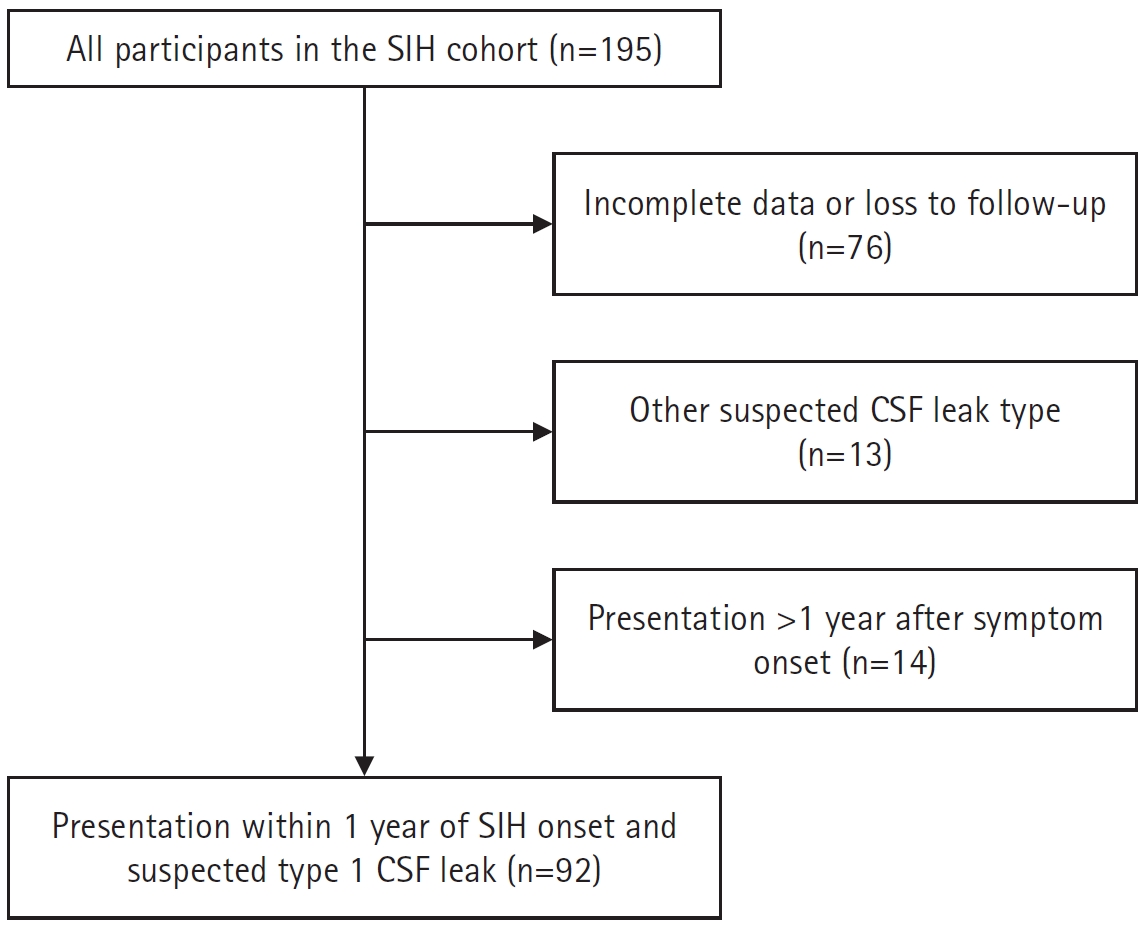
Study flowchart. Of the 195 patients in the SIH cohort, 76 were excluded because of incomplete data or loss to follow-up, 13 because of other suspected CSF leak types, and 14 because they presented more than 1 year after symptom onset. The final study population consisted of 92 patients with a suspected type 1 CSF leak who presented within 1 year of SIH onset.
SIH, spontaneous intracranial hypotension; CSF, cerebrospinal fluid.
2. Participants
All patients met the diagnostic criteria for SIH according to the International Classification of Headache Disorders, 3rd edition,11 and were diagnosed in person by a board-certified neurologist with expertise in headache and SIH. In this study, we enrolled patients in whom a type 1 leak was suspected based on the spinal magnetic resonance myelogram (MRM) findings. Patients with CSF leakage attributable to lumbar puncture, another spinal procedure, or trauma were excluded in accordance with the International Classification of Headache Disorders, 3rd for SIH. The analysis was restricted to patients presenting within 1 year of symptom onset to reduce potential selection bias, as patients presenting after a longer disease duration may represent a selected population with persistent or more severe symptoms. Among 195 patients enrolled to the SIH registry, 92 met the inclusion criteria.
Demographic and clinical variables including age, sex, symptom onset, follow-up duration, time from onset to first epidural blood patch (EBP), number of EBPs administered, and brain magnetic resonance imaging (MRI) findings (including brain sagging, pachymeningeal enhancement, venous sinus engorgement, pituitary hyperemia, subdural hygroma, subdural hematoma, and cerebral venous thrombosis) were prospectively collected.
All patients underwent brain MRI and noninvasive MRM using a standardized protocol, which included fat-saturated T2-weighted imaging, heavily T2-weighted 3-dimensional sequences and multi-planar sagittal and axial reconstructions using water-only and in-phase T2-weighted images. Follow-up MRM was primarily performed after EBP to assess treatment response or when deemed clinically necessary. Because follow-up MRM was optional and non-reimbursed, the timing and frequency of imaging varied among patients. Ultrafast dynamic computed tomography myelography (CTM) was performed in selected cases.
3. Outcome evaluation and definition
The primary outcome was incident SCH, defined as the first radiologically confirmed event following SIH diagnosis. All participants had no evidence of SCH at baseline. Serial imaging was reviewed to confirm that no baseline protrusion was present at the level of SCH, thus SCH represented a newly developed lesion at a previously uninvolved spinal level.
Radiological diagnosis of SCH was defined as a focal protrusion of the spinal cord beyond the expected ventral dural margin, showing an identifiable point where the cord extends outside the dural sac, while maintaining continuity within the sac at adjacent levels. The lesion was identified on both sagittal and axial MRM. In patients with suspected SCH on MRM, ultrafast dynamic CTM was also used for further evaluation and confirmation.
Every case was reviewed by a senior headache neurologist and SIH specialist (MJL, with 16 years of experience in neurology and headache) and two senior radiologists (HJY and JYC, with 21 and 27 years of experience in musculoskeletal imaging, respectively). SCH was diagnosed only in cases where all of them agreed on the diagnosis.
4. Statistical analysis
Kaplan–Meier survival analysis was performed to estimate the cumulative incidence of SCH over time, with an event defined as the first confirmed occurrence of SCH. Censoring was defined as the duration from the onset to the last follow-up date for those without events and the last MRM date for those who achieved resolution in the non-SCH group. In patients with SCH, the duration from symptom onset to SCH occurrence was measured. As a sensitivity analysis, we repeated the Kaplan–Meier analysis using a delayed-entry approach, with symptom onset as the underlying time scale and enrollment as the time of entry into the risk set. Patients entered the risk set at enrollment and remained at risk until SCH occurrence or the censoring time defined in the primary analysis.
Cox proportional hazards regression analysis was performed to evaluate the effects of demographic and clinical variables on the risk of developing SCH. The results are presented as hazard ratios (HRs) with 95% confidence intervals (CIs). The incidence rates were calculated based on the total person-years of follow-up. Statistical significance was defined as a two-sided p<0.05. All analyses were performed using the R software version 4.3.3.
RESULTS
1. Baseline characteristics of study participants
Of the 195 registered patients with SIH, 92 with SLEC were considered eligible and included in the analysis (Table 1). The mean age was 40.6±10.7 years, with 39.1% male. The mean disease duration from onset to the first visit was 54.4±63.7 days, and patients underwent 3.8±2.0 MRM follow-ups for 301.8±271.7 days. Included patients received an average of 4.6±2.6 EBPs. Brain MRI abnormalities were observed in 52 patients (56.5%), most commonly pachymeningeal enhancement (32.6%), venous sinus engorgement (32.6%), and subdural hematoma (19.6%). No patients exhibited spinal cord herniation at baseline.
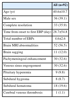
Demographic and clinical characteristics of the study participants
2. Spinal cord herniation incidence
Among the 92 patients who presented within 1 year of SIH onset and had SLEC with suspected type 1 CSF leakage, 8 (8.7%) developed SCH during the prospective observation over 405.5±275.4 days. Baseline spinal MRM was obtained at 17, 77, 10, 19, 33, 3, 59, and 8 days after symptom onset, respectively, and SCH was first detected on follow-up MRM at 328, 547, 145, 71, 174, 34, 78, and 16 days, respectively. Baseline MRM showed no SCH at the subsequently affected level in any patient. Seven of the eight cases were detected within 1 year and all were detected within 2 years.
The number of at-risk patients progressively decreased over time (Figure 2). By 90 days, four SCH events had occurred, resulting in an incidence of 4.9% (95% CI, 0.09%–9.56%). At 180 days, the cumulative number of SCH events increased to six, raising the incidence to 8.2% (95% CI, 1.60%–14.36%). By day 270, the cumulative number of events remained at six. At 1 year, the cumulative number of SCH events increased to seven, corresponding to an incidence of 10.3% (95% CI, 2.54%–17.41%). Finally, by 2 years, the cumulative number of SCH events reached eight, with an incidence of 14.2% (95% CI, 3.23%–23.90%). The overall SCH incidence during the study period was 0.092 per person-years, calculated over a total follow-up time of 86.95 person-years.
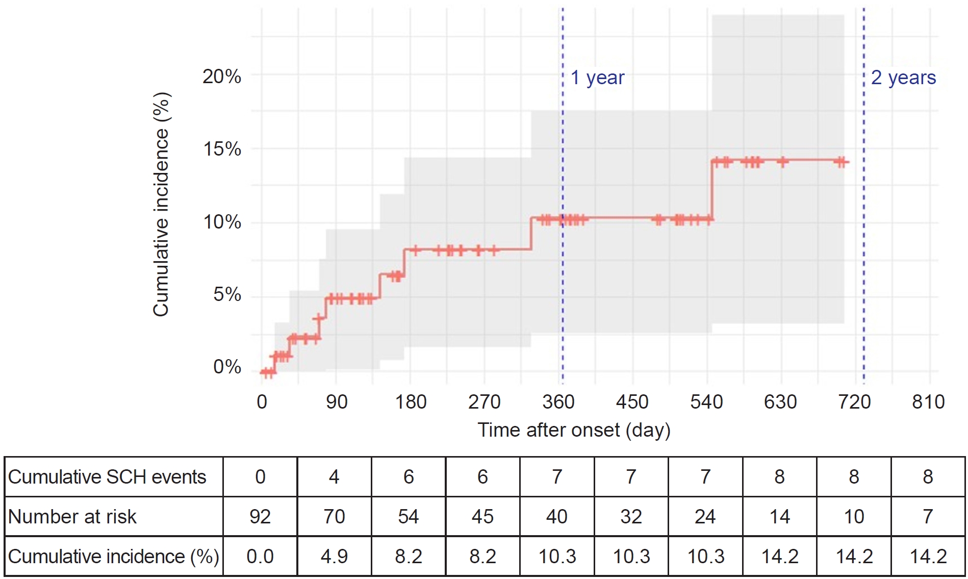
Kaplan–Meier curve for spinal cord herniation. The Kaplan–Meier curve estimates the cumulative incidence of spinal cord herniation (SCH). The x-axis shows the time in days from symptom onset to SCH development or censoring; “+” marks indicate censoring. Time is shown in 90-day increments to illustrate early SCH development in greater detail. The table beneath the x-axis lists the cumulative number of SCH events, the number of patients at risk, and the incidence. The y-axis shows the cumulative incidence of SCH. The gray shading indicates the 95% confidence intervals, which widened as the number of patients at risk decreased, reflecting greater variability in the estimate.
In the sensitivity analysis accounting for delayed entry, four patients could not contribute to the analysis because they underwent EBP on the day of enrollment and SLEC resolution was confirmed on post-EBP MRM performed the same day, resulting in no post-entry observation time. Thus, the analysis included 88 patients and all eight SCH events. The estimated cumulative incidence of SCH was 16.8% (95% CI, 2.9%–28.8%) at 1 year and 20.5% (95% CI, 5.0%–33.4%) at 2 years.
3. Factors associated with spinal cord herniation
Comparison between patients with and without herniation revealed a male predominance in the herniation group (87.5% vs. 34.5%, p=0.005) and a shorter interval from symptom onset to the first EBP (p=0.016) (Table 2). No significant differences were observed in age, follow-up duration, total number of EBPs, or brain MRI findings between the groups. In univariable Cox regression analysis, male sex was associated with SCH development (HR, 13.35; 95% CI, 1.64–109.00; p=0.016) (Table 3).
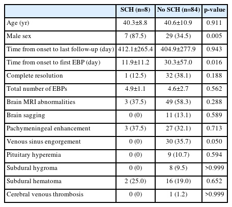
Comparison of baseline characteristics between patients with and without SCH
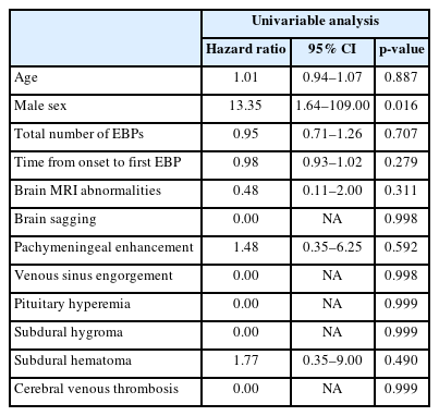
Factors associated with spinal cord herniation
4. Clinical and radiologic characteristics of participants with spinal cord herniation
Among the eight patients who developed SCH, the time from symptom onset to initial detection ranged from 16 to 547 days. Seven patients were identified within the first year and the remaining patient between 1 and 2 years. SCH was detected after a mean of 3.1±2.36 follow-up MRM examinations. None of the patients had neurological symptoms attributable to spinal cord compression or irritation at the time of SCH detection. Radiologic findings of SCH on MRM and ultrafast dynamic CTM are shown in Figures 3 and 4, respectively. Following SCH progression, improvement in orthostatic headache was observed in three patients (37.5%), reduction in the extent of SLEC in four patients (50.0%), and false-negative ultrafast dynamic CTM findings in three patients (37.5%) paradoxically. Intraoperative findings demonstrating SCH and the corresponding ventral dural defects are shown in Figure 5.
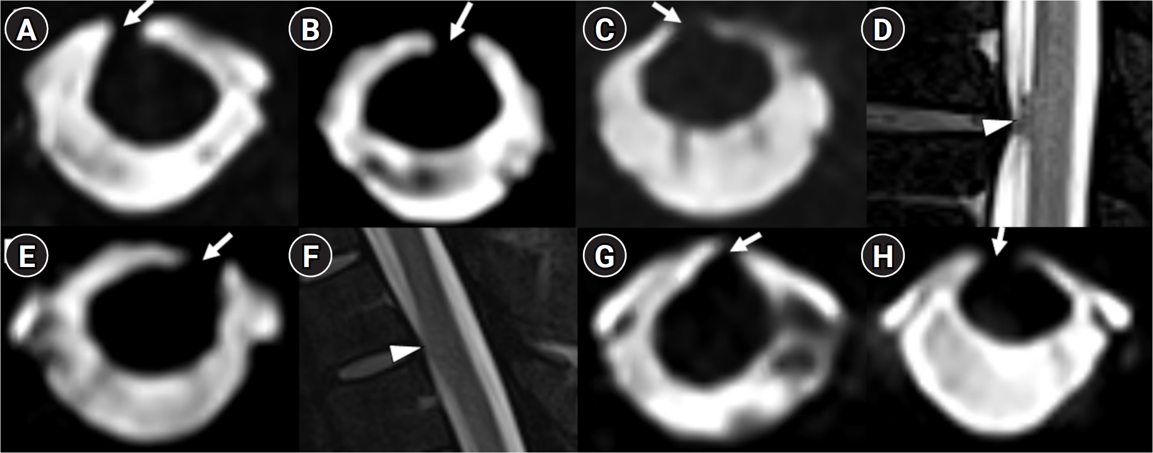
Spinal cord herniation on magnetic resonance myelography. (A–C, E, G, H) Axial T2-weighted magnetic resonance myelograms demonstrating spinal cord herniation (arrows). (D, F) Sagittal T2-weighted magnetic resonance myelograms demonstrating spinal cord herniation (arrowheads).
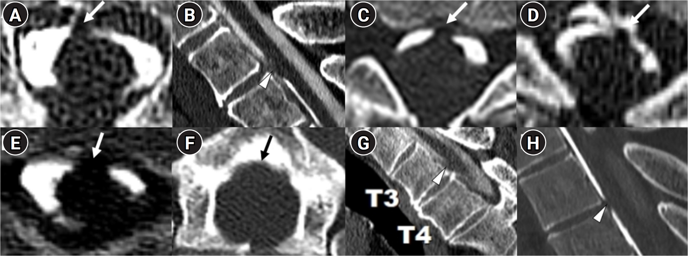
Spinal cord herniation on ultrafast computed tomography myelography. (A, C–F) Axial ultrafast computed tomography myelograms demonstrating spinal cord herniation (arrows). (B, G, H) Sagittal ultrafast computed tomography myelograms demonstrating spinal cord herniation (arrowheads).
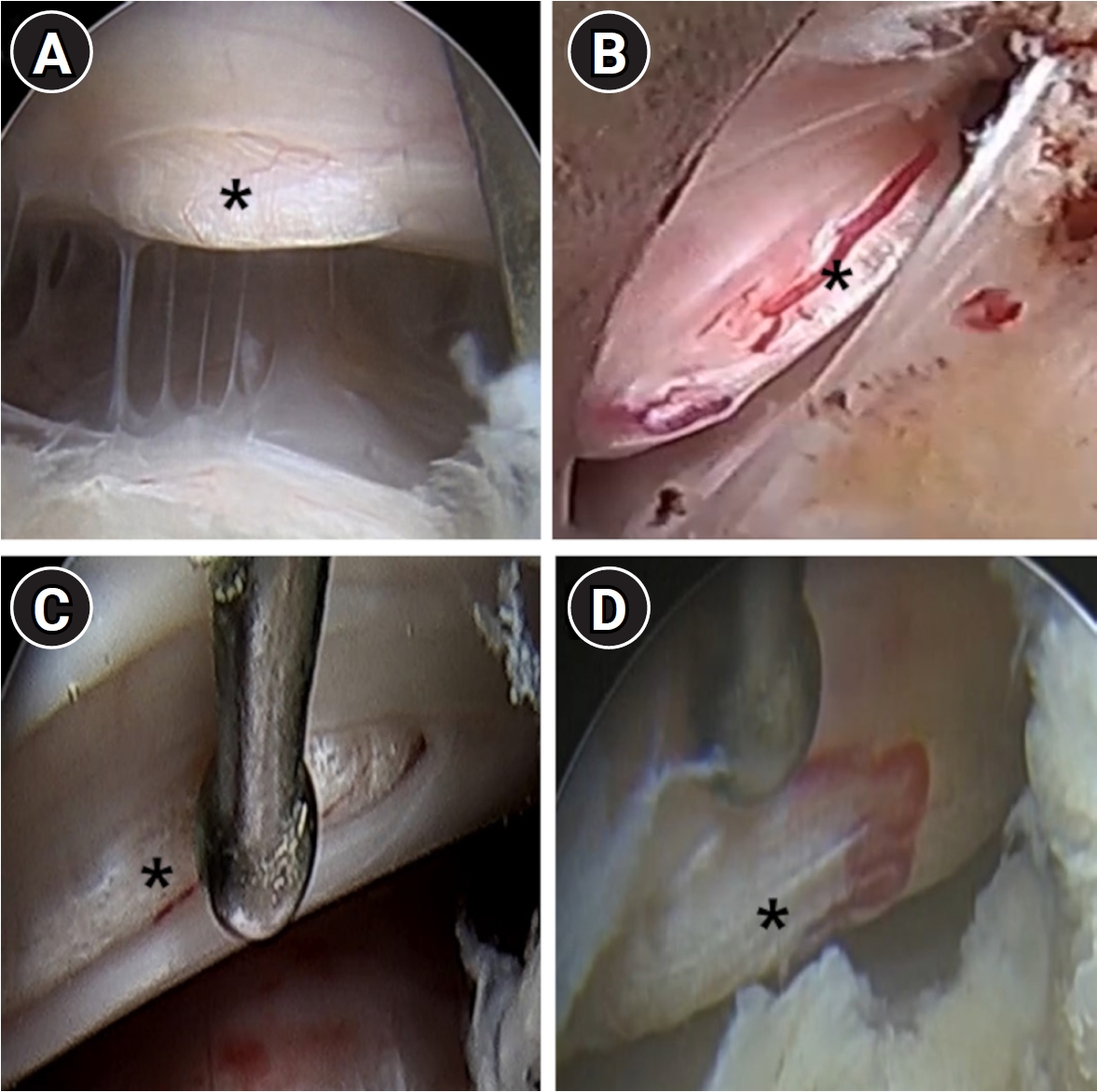
Intraoperative findings in patients with spinal cord herniation. (A) In a 36-year-old woman, surgery 760 days after symptom onset revealed spinal cord herniation (asterisk) through dural tears. (B) In a 48-year-old man, surgery 340 days after symptom onset revealed spinal cord herniation (asterisk) through dural tears. (C) In a 33-year-old man, spinal cord herniation (asterisk) through dural tears was confirmed intraoperatively 174 days after onset. (D) In a 52-year-old man, surgery 103 days after symptom onset revealed spinal cord herniation (asterisk) through dural tears.
DISCUSSION
The major findings of the study are as follows. First, to the best of our knowledge, this is the first report to document the early development of SCH in patients with SIH. Second, SCH occurs more frequently and rapidly than previously expected. Third, early SCH showed distinct clinical and radiological features.
This is the first report on the early development of SCH in patients with SIH. SCH has been considered a chronic, yet rare, complication of SIH. In our cohort of 92 patients who presented within 1 year of SIH onset and had SLEC with suspected type 1 CSF leakage, SCH developed in eight patients, yielding a cumulative incidence of approximately 10.3% by 1 year and 14.2% by 2 years. This finding contrasts with the only other report on SCH, which was conducted by a neurosurgical group in the United States. In that study, only 1 of 1,589 patients with SIH developed SCH over a 20-year period.4 In a subsequent analysis of 51 patients with persistent ventral CSF leaks within 1 year from symptom onset and followed up for >1 year without surgery, no cases of SCH were initially identified. However, during long-term follow-up, SCH eventually developed in two of these 51 patients, with a reported cumulative incidence of 0% at 5 years and 9.4% at 10 years.8,10 By contrast, SCH in our study developed within a significantly shorter timeframe, with most cases occurring before 2 years.
This discrepancy, particularly the strikingly high incidence rate of SCH in our study, can be explained by the differences in clinical settings. In South Korea, the surgical treatment of ventral spinal dural defects was almost unavailable before October 2024. As a result, even refractory patients requiring surgery were managed with EBPs alone. This allowed us to longitudinally observe the clinical evolution of refractory persistent ventral CSF leaks. In contrast, the only other report on SCH involved a neurosurgical group where surgery was actively available. Their analysis was limited to patients with ventral dural defects who did not undergo surgery, as patients with more severe or refractory disease were more likely to receive definitive surgical treatment. Consequently, their cohort likely represented a relatively milder subset of patients.8,10 Moreover, in previous studies, surgical treatment was conducted with a posterior approach via laminectomy, which may be less ideal to detect spinal cord protrusion to the ventral epidural space.12 In contrast, we were able to directly visualize and identify SCH more accurately by utilizing anterior endoscopic approaches. Another possible explanation is that patients underwent multiple MRMs (mean, 5.5±4.68 times/yr) in our study. Repeated MRM may have increased the likelihood of detecting early asymptomatic SCH, which may partly explain the earlier and higher observed incidence compared with previous studies. However, repeated imaging allowed detection of SCH that might otherwise have remained unrecognized with less frequent imaging. Thus, serial imaging may facilitate the detection of SCH before the development of neurological symptoms. These fundamental differences may explain the higher incidence of SCH reported in our study.
Several clinical characteristics of early SCH warrant attention. Male sex was associated with SCH development in the univariable analysis; however, given the small number of events and the resulting imprecision of the estimate, this finding should be considered exploratory. Sex-related differences in ventral spinal structures, including calcification or bony spurs and more repetitive mechanical friction related to spinal mobility, may potentially influence susceptibility to SCH. Notably, none had neurological symptoms attributable to spinal cord compression or irritation at the time of SCH detection, suggesting that early SCH may remain neurologically silent. In addition, some patients exhibited paradoxical clinical or radiological improvement following SCH development, including improvement in orthostatic headache, reduction in SLEC, or absence of detectable CSF leakage on dynamic CTM. These changes may represent “pseudo-improvement” resulting from partial obstruction of the dural defect by the herniated spinal cord, a phenomenon that is being investigated in greater detail in a separate study. Thus, early SCH may remain clinically inconspicuous and should not be overlooked, particularly in the absence of neurological deficits or when manifestations appear to improve.
Our study has several strengths. The limited availability of definitive surgical treatment during most of the study period provided a unique opportunity to longitudinally observe the development of SCH in patients with persistent ventral CSF leaks. In addition, prospectively collected clinical data and serial spinal imaging enabled characterization of the timing of newly developed SCH. However, several limitations should be acknowledged. First, this was a single-center tertiary referral cohort specializing in CSF leak disorders. Patients referred to our center are likely to represent more severe or persistent disease, and the study population was further restricted to patients presenting within 1 year of symptom onset with SLEC suggestive of a type 1 CSF leak. These referral and selection factors may have contributed to the relatively high observed incidence of SCH. Therefore, the cumulative incidence estimates should be interpreted as specific to this selected cohort and should not be generalized to the overall SIH population. Second, repeated EBPs during follow-up may have influenced the natural course of CSF leakage and SCH development. Third, the timing of SCH was defined by its first radiological detection. Because imaging was performed at discrete follow-up intervals, the true onset of SCH may have preceded its radiological diagnosis, resulting in uncertainty in time-to-event estimation. Because some patients underwent spinal imaging before referral, institutional enrollment did not necessarily represent the start of radiological observation. We therefore retained the original analysis as the primary descriptive analysis of the full cohort. However, including pre-enrollment person-time may introduce bias, and the higher estimates obtained in the delayed-entry analysis indicate that the absolute cumulative incidence estimates depend on the handling of entry time and should be interpreted cautiously. In addition, frequent MRM follow-up may have introduced ascertainment bias. However, more frequent imaging may have facilitated earlier detection of asymptomatic SCH, whereas less frequent imaging may have resulted in underestimation of its incidence. Finally, the small number of SCH cases limited the precision of the incidence and risk estimates, particularly the observed association with male sex.
CONCLUSION
Among patients presenting within 1 year of symptom onset with SLEC suggestive of a type 1 CSF leak, SCH may develop earlier than previously recognized, even in the absence of neurological symptoms. Careful surveillance may facilitate early detection and appropriate management. However, the incidence estimate should be interpreted with caution when generalizing to the overall SIH population.
Notes
AVAILABILITY OF DATA AND MATERIAL
The data used in this study included sensitive patient information and were protected under the institutional review board guidelines. Therefore, raw data cannot be made publicly available or shared upon request due to ethical concerns. All data and materials supporting the conclusions of this study have been included in this article.
AUTHOR CONTRIBUTIONS
Conceptualization: SAK, MJL; Data curation: SAK, SC, KY, HJY, JYC, MJL; Formal analysis: SAK, MJL; Investigation: SAK, SC, KY, HJY, JYC, MJL; Methodology: SAK, MJL; Visualization: SAK, MJL; Writing–original draft: SAK, MJL; Writing–review & editing: SAK, SC, KY, HJY, JYC, MJL.
CONFLICT OF INTEREST
No potential conflict of interest relevant to this article was reported.
FUNDING STATEMENT
This work was supported by a National Research Foundation of Korea grant funded by the Korean government (RS-2024-00357394) and a Korea Medical Device Development Fund grant funded by the Korean government (Ministry of Science and ICT, Ministry of Trade, Industry and Energy, Ministry of Health & Welfare, Ministry of Food and Drug Safety) (project number: RS-2023-00229484).
ACKNOWLEDGMENTS
None.